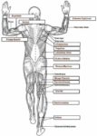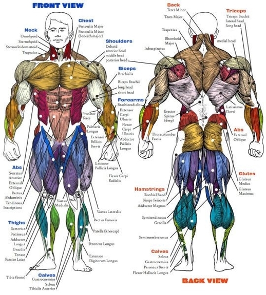
Human Muscle Structure
The human muscle system is a complex network of tissues designed to provide movement and maintain posture. Broadly, human muscles can be classified into three types: striated (or skeletal) muscle, smooth muscle, and cardiac muscle.
1. Striated (Skeletal) Muscle: These muscles are attached to the bones by tendons and are under voluntary control. They are responsible for all locomotion and mechanical bodily functions. For example, the biceps brachii muscle enables the bending of the elbow. There are more than 600 skeletal muscles in the human body, making up about 40% of a person’s body weight. Each skeletal muscle is a discrete organ constructed of muscle tissue, blood vessels, tendons, and nerves.
2. Smooth Muscle: Found in the walls of structures such as the urinary bladder, intestines, stomach, respiratory passageways, and blood vessels. These muscles are under involuntary control and their contractions are responsible for the wavelike movements that propel substances through the bodily system.
3. Cardiac Muscle: This muscle type makes up the mass of the heart and is responsible for the rhythmic contractions of this vital pumping organ. It is under involuntary control and contracts in response to signals from the brain.
Each muscle consists of fibers of muscle cells surrounded by protective tissue. Bundled together are many more fibers, all surrounded by a thick protective tissue. Each fiber comprises many tiny strands called fibrils, and impulses from nerve cells control the contraction of each muscle fiber.
Muscle movement happens when neurological signals produce electrical changes in muscle cells. During this process, calcium is released into the cells and brings about a short muscle twitch. Problems with the junction between the cells, called a synapse, can lead to neuromuscular diseases.
Proper nutrition and exercise are important for keeping all muscles healthy, whether they are cardiac, smooth, or skeletal. Some muscular disorders and conditions that affect muscles include muscle pain, sprains and strains, bruising, cramping, myopathy, muscular dystrophy, Parkinsons disease, fibromyalgia, and multiple sclerosis.
In conclusion, the human muscle system is a marvel of biological engineering, enabling us to perform a vast range of movements and tasks. Understanding its structure and function is crucial for maintaining health and treating diseases.