Human Heart Anatomy
The human heart, a muscular organ, is the center of the circulatory system. It is located between the lungs, slightly to the left of center, behind the breastbone, and rests on the diaphragm. The heart is approximately the size of a closed fist.
tructure
The heart consists of several layers of a tough muscular wall, known as the myocardium. A thin layer of tissue, the pericardium, covers the outside, and another layer, the endocardium, lines the inside.
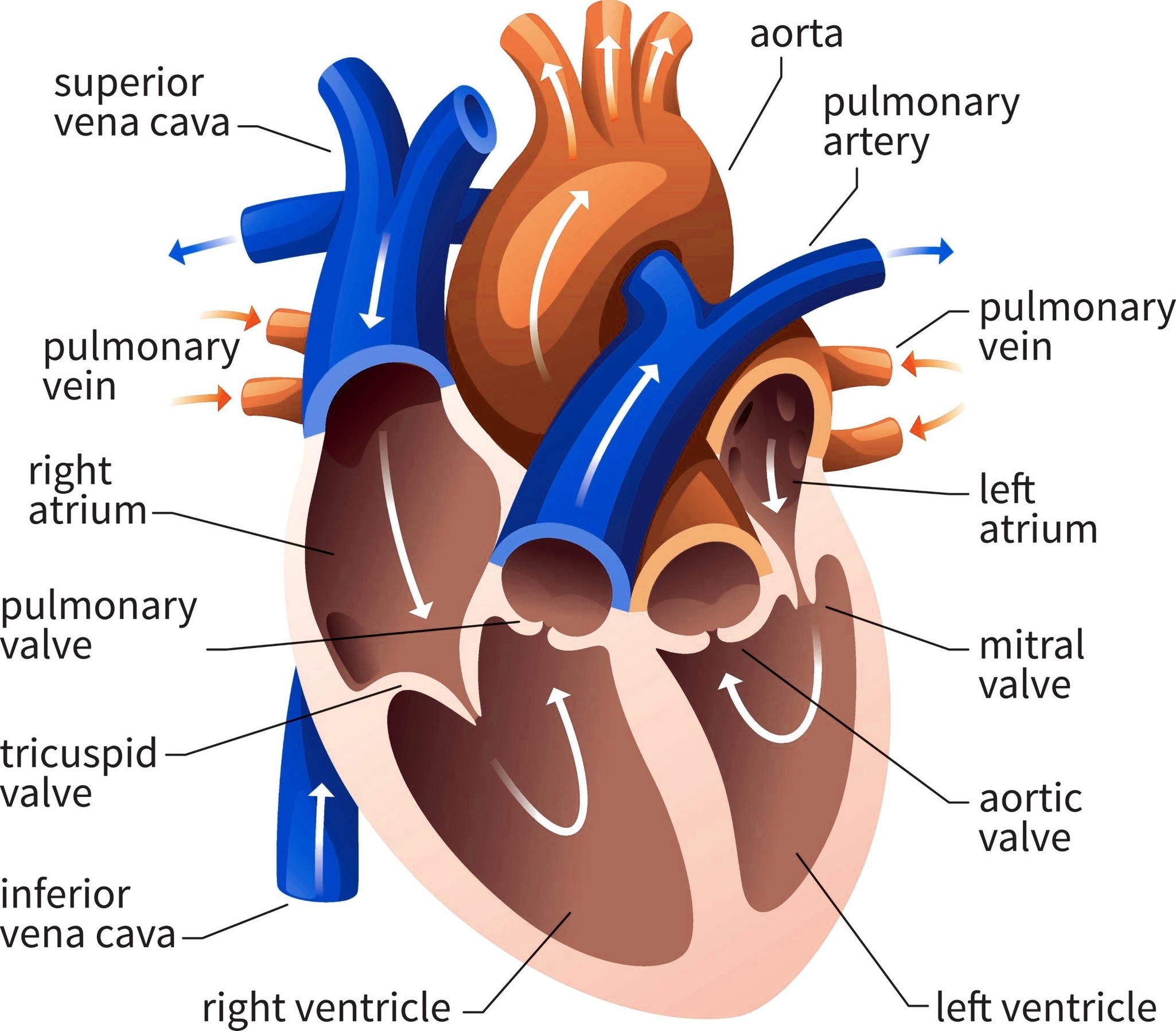
The heart cavity is divided down the middle into a right and a left heart, each subdivided into two chambers. The upper chamber is called an atrium (or auricle), and the lower chamber is called a ventricle.
Chambers and Valves
The two atria act as receiving chambers for blood entering the heart, while the more muscular ventricles pump the blood out of the heart. The right atrium receives venous blood from the head, chest, and arms via the superior vena cava, and from the abdomen, pelvic region, and legs via the inferior vena cava.
Blood then passes through the tricuspid valve to the right ventricle, which propels it through the pulmonary artery to the lungs. The left atrium and ventricle receive oxygenated blood from the lungs and pump it to the systemic vessels, which distribute it throughout the body.
Function
The heart, although a single organ, can be considered as two pumps that propel blood through two different circuits. The right side of the heart receives deoxygenated blood from the body and pumps it to the lungs for oxygenation. The left side of the heart receives the oxygenated blood from the lungs and pumps it to the rest of the body.
Coronary Circulation
The heart’s own blood supply is provided by the coronary arteries. The right coronary artery supplies the right atrium, right ventricle, and parts of the left ventricle and septum. The left coronary artery supplies the left atrium