Human Heart Anatomy
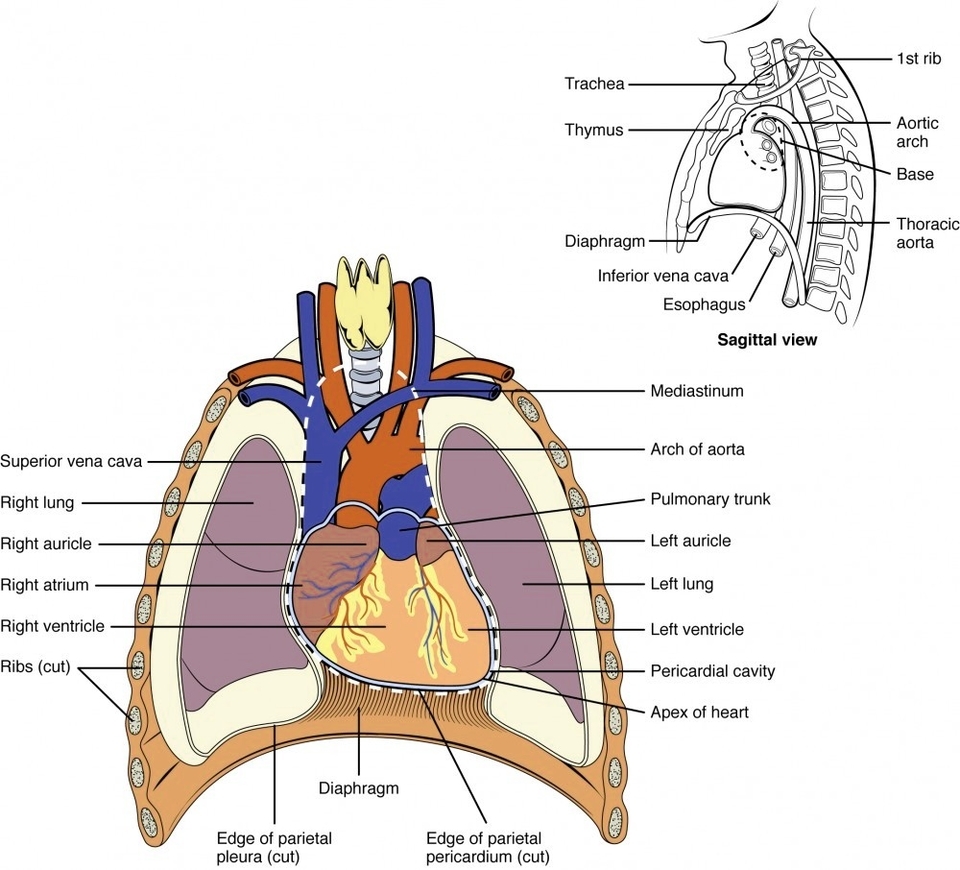
The human heart is a muscular organ that plays a vital role in the circulatory system. It pumps oxygenated blood throughout the body and deoxygenated blood to the lungs. The heart is located in the middle mediastinum, enclosed within a two-layered serous sac called the pericardium. It is shaped like a quadrangular pyramid, with its base facing the posterior thoracic wall and its apex pointed towards the anterior thoracic wall.
Heart Structure
The heart is divided into four chambers: two atria (right and left) and two ventricles (right and left). The right atrium and ventricle receive deoxygenated blood from systemic veins and pump it to the lungs, while the left atrium and ventricle receive oxygenated blood from the lungs and pump it to the systemic vessels, which distribute it throughout the body.
Heart Wall
The heart wall is composed of three layers:
1. Epicardium: The outermost layer, also known as the visceral pericardium, covers the heart and adheres the heart wall to a protective sac.
2. Myocardium: The middle layer, composed of strong muscle tissue, powers the heart’s pumping action.
3. Endocardium: The innermost layer, lines the interior structures of the heart.
Heart Valves
Four valves regulate and support the flow of blood through and out of the heart. Each valve is formed by a group of folds, or cusps, that open and close as the heart contracts and dilates. The valves include:
1. Tricuspid Valve: Located between the right atrium and the right ventricle.
2. Pulmonary Valve: Manages blood flow out of the right ventricle through the pulmonary trunk into the pulmonary arteries.
3. Mitral Valve: Located between the left atrium and the left ventricle.
4. Aortic Valve: Manages blood flow from the left ventricle into the aorta.
Coronary Circulation
The heart receives its blood supply from the right and left coronary arteries. The right coronary artery has several branches, including the sinuatrial nodal branch, right marginal branch, atrioventricular nodal branch, and posterior interventricular branch. The left coronary artery has the circumflex branch and the anterior interventricular branch.
Conclusion
The human heart, with its intricate structure and complex functions, is a marvel of biological engineering. Its ceaseless work keeps us alive, circulating oxygen-rich blood throughout our bodies and returning deoxygenated blood to the lungs for reoxygenation. Understanding the anatomy of the heart is crucial for comprehending many aspects of human health and disease..