Human Heart Anatomy
The human heart, a muscular organ located between the lungs in the middle compartment of the chest, is the center of the circulatory system. It’s approximately the size of a closed fist and is shaped like a quadrangular pyramid. The heart’s primary function is to pump blood throughout the body via the circulatory system.
tructural Composition
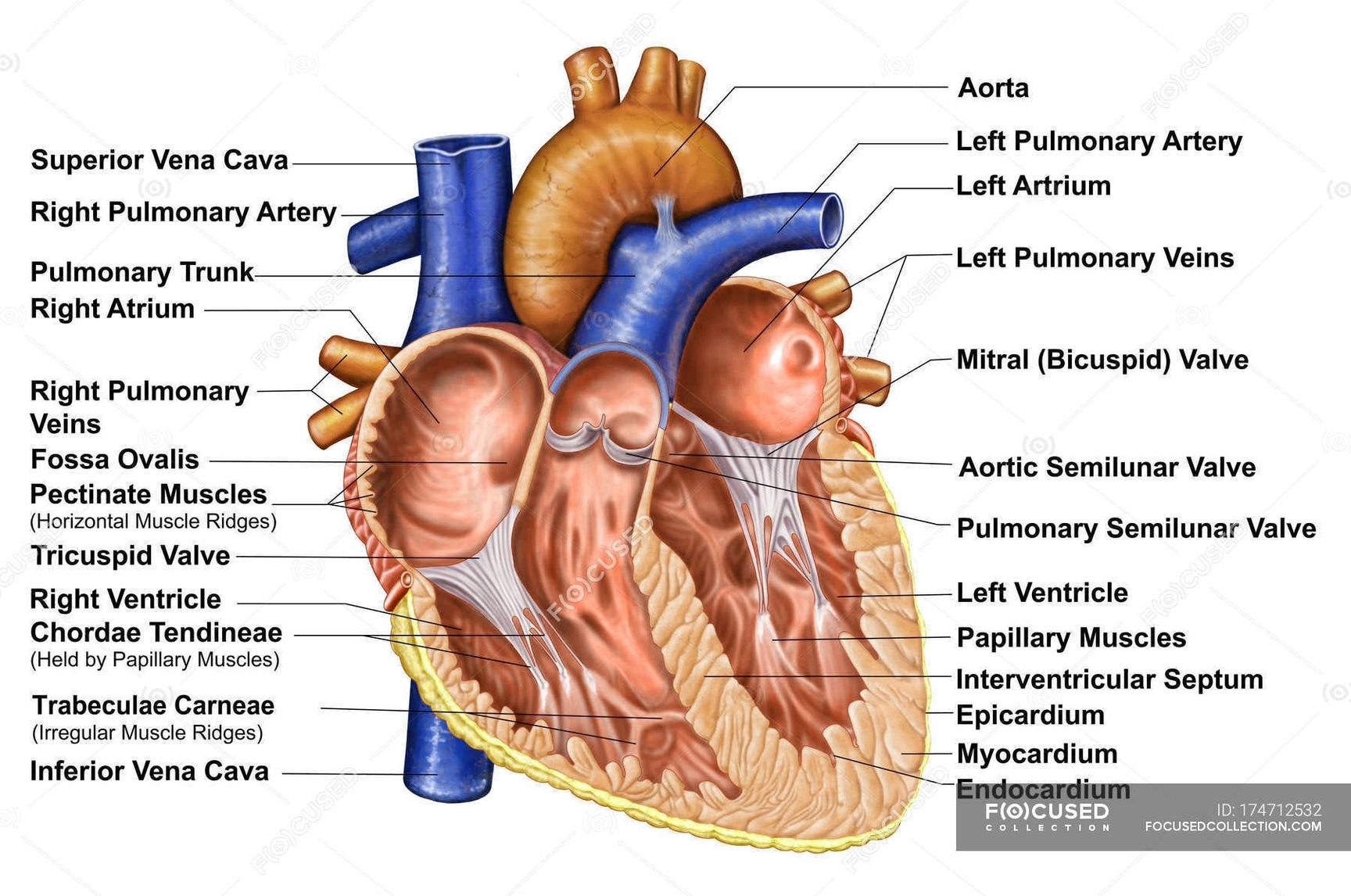
The heart consists of several layers of a tough muscular wall, known as the myocardium. A thin layer of tissue, the pericardium, covers the outside, and another layer, the endocardium, lines the inside.
Chambers and Valves
The heart is divided into a right and a left heart, each subdivided into two chambers: an upper chamber called an atrium (or auricle) and a lower chamber called a ventricle. The two atria act as receiving chambers for blood entering the heart, while the more muscular ventricles pump the blood out of the heart.
The heart has four valves that ensure blood flows in the right direction. These are the tricuspid, pulmonary, mitral, and aortic valves.
Circulatory Function
The heart can be considered as two pumps that propel blood through two different circuits. The right atrium receives venous blood from the head, chest, and arms via the superior vena cava, and from the abdomen, pelvic region, and legs via the inferior vena cava. Blood then passes through the tricuspid valve to the right ventricle, which propels it through the pulmonary artery to the lungs.
Coronary Circulation
The heart receives its own supply of blood from the coronary arteries. Two major coronary arteries branch off from the aorta near the point where the aorta and the left ventricle meet.
Conclusion
The human heart, with its intricate structure and complex functions, is a marvel of biological engineering. Its ceaseless work keeps us alive, circulating oxygen-rich blood throughout our bodies. Understanding its anatomy and function is fundamental to comprehending many aspects of human health and disease..