Human Ear Anatomy
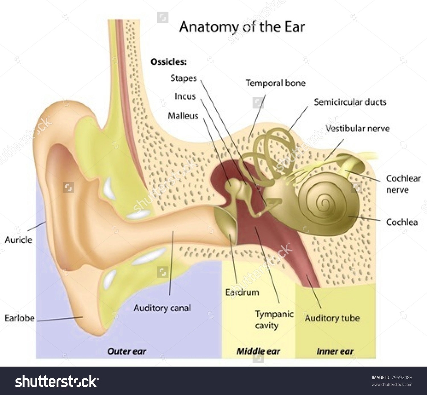
The human ear is a complex sensory organ responsible for hearing and maintaining balance. It is anatomically divided into three parts: the outer ear, middle ear, and inner ear.
Outer Ear
The outer ear consists of the visible portion called the auricle or pinna, which projects from the side of the head, and the short external auditory canal. The inner end of the canal is closed by the tympanic membrane, commonly known as the eardrum. The outer ear’s function is to collect sound waves and guide them to the tympanic membrane.
Middle Ear
The middle ear is a narrow air-filled cavity in the temporal bone. It is spanned by a chain of three tiny bones the malleus (hammer), incus (anvil), and stapes (stirrup), collectively known as the auditory ossicles. This ossicular chain conducts sound from the tympanic membrane to the inner ear.
Inner Ear
The inner ear, known since the time of Galen (2nd century CE) as the labyrinth, is a complicated system of fluid-filled passages and cavities located deep within the rock-hard petrous portion of the temporal bone. It consists of two functional units: the vestibular apparatus, consisting of the vestibule and semicircular canals, which contains the sensory organs of postural equilibrium; and the snail-shell-like cochlea, which contains the sensory organ of hearing. These sensory organs are highly specialized endings of the eighth cranial nerve, also called the vestibulocochlear nerve.
Function
The main functions of the ear are hearing and maintaining balance. The eardrum vibrates from the incoming sound waves and sends these vibrations to the three tiny bones in the middle ear. These bones amplify the sound vibrations and send them to the cochlea, a snail-shaped structure filled with fluid, in the inner ear. The cochlea then transforms these vibrations into electrochemical impulses that are sent to the brain via the vestibulocochlear nerve.
Conclusion
The human ear is a marvel of biological engineering, capable


