The right atrium is one of the four chambers of the heart. The heart is comprised of two atria and two ventricles. Blood enters the heart through the two atria and exits through the two ventricles. Deoxygenated blood enters the right atrium through the inferior and superior vena cava.
On frontal CXR the findings are consistent with a large right atrium characterized by the globular shape of the heart, large right heart border occupying more than 50% of the cardiovascular height. DOMINANT RIGHT ATRIAL ENLARGEMENT.
On frontal CXR the findings are consistent with a large right atrium characterized by the globular shape of the heart, large right heart border occupying more than 50% of the cardiovascular height. DOMINANT RIGHT ATRIAL ENLARGEMENT.
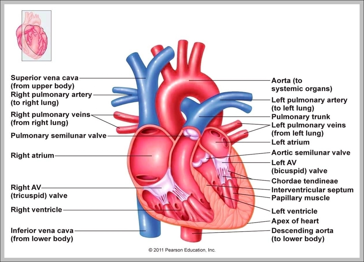
Heart Right Atrium Image
Posted inDiagrams
Heart Right Atrium Image
Post navigation
Previous Post
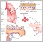 Function Of The Lungs Image
Next Post
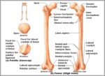
Anatomy Of Femur Image