The femur, also known as the thigh bone, is the longest and strongest bone in the human body. It plays a crucial role in supporting body weight and facilitating movement. The femur can be divided into three main parts: the proximal end, the shaft, and the distal end.
Proximal End
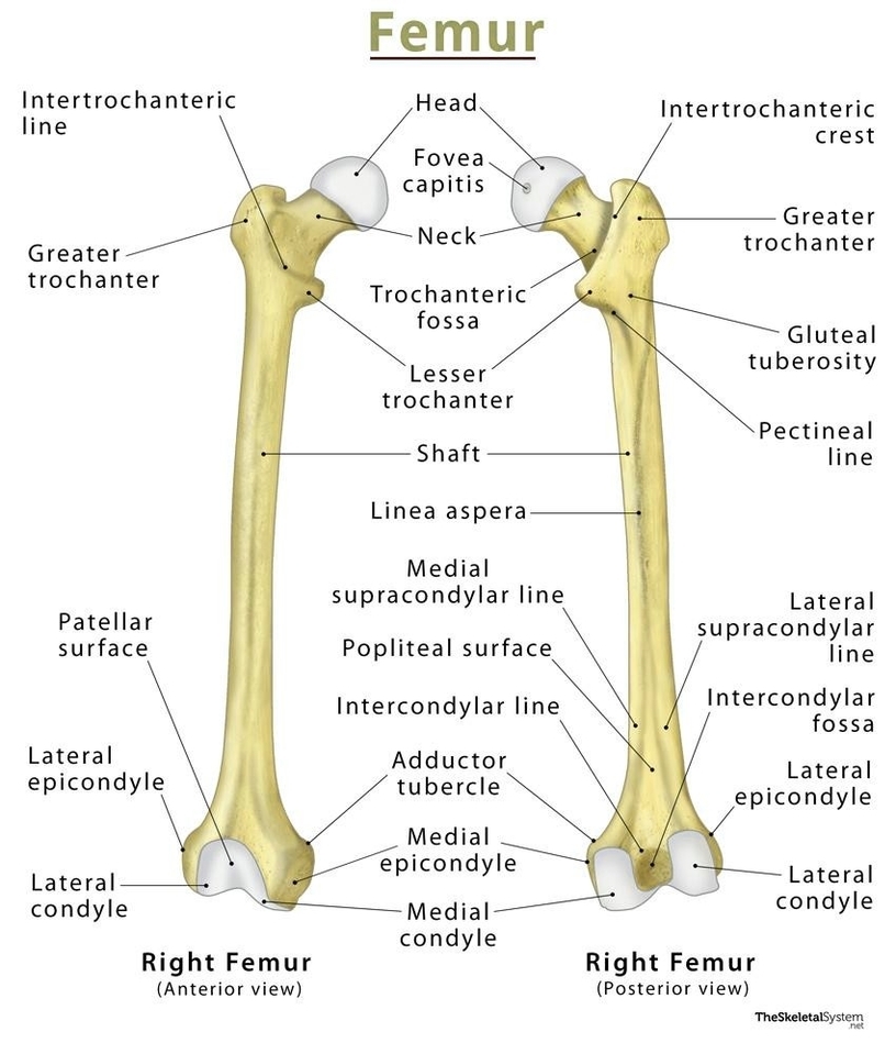
The proximal end of the femur articulates with the acetabulum of the pelvis to form the hip joint. It consists of the head, neck, and two bony processes the greater and lesser trochanters. The head of the femur is a roughly spherical structure that sits superomedially and projects anteriorly from the neck of the femur. The smooth convexity of the femoral head is disrupted on the posteroinferior surface by a depression known as the fovea for the ligament of the head (fovea capitis femoris).
haft
The shaft of the femur is the long, cylindrical part of the bone. It has spongy bone tissue at both ends and a cavity filled with bone marrow in the shaft. The shaft has several borders and surfaces, including lateral and medial borders, and anterior, medial, and lateral surfaces. It also features several ridges, such as the lateral ridge (gluteal tuberosity), pectineal line, and spiral line, which converge to form the linea aspera.
Distal End
The distal end of the femur articulates with the tibia to form the knee joint. It consists of the lateral and medial condyles, intercondylar fossa, and lateral and medial epicondyles.
Blood Supply
The femur receives blood supply from the trochanteric anastomosis and cruciate anastomosis.
Muscle Attachments
The femur serves as the site of origin and attachment for many muscles and ligaments. For instance, the greater trochanter is the site of attachment for many of the muscles in the gluteal region, such as gluteus medius, gluteus minimus, and piriformis. The lesser trochanter is the site of attachment for the iliopsoas muscle.
Development
The femur begins to develop between the 5th to 6th gestational week by way of endochondral ossification (where a bone is formed using a cartilage-based foundation). While several ossification centers (points of bone development) appear throughout intrauterine life, the bone continues to develop through childhood and early adolescence. Ossification of the femur is completed between the 14th and 18th years of life.
Disorders
There are several disorders associated with the femur, including neck of femur fractures, slipped capital femoral epiphysis, and femoroacetabular impingement.
In conclusion, the femur is a vital bone in the human body, playing a key role in locomotion and weight-bearing. Its unique anatomy makes it suitable for supporting the numerous muscular and ligamentous attachments within the lower limb region..