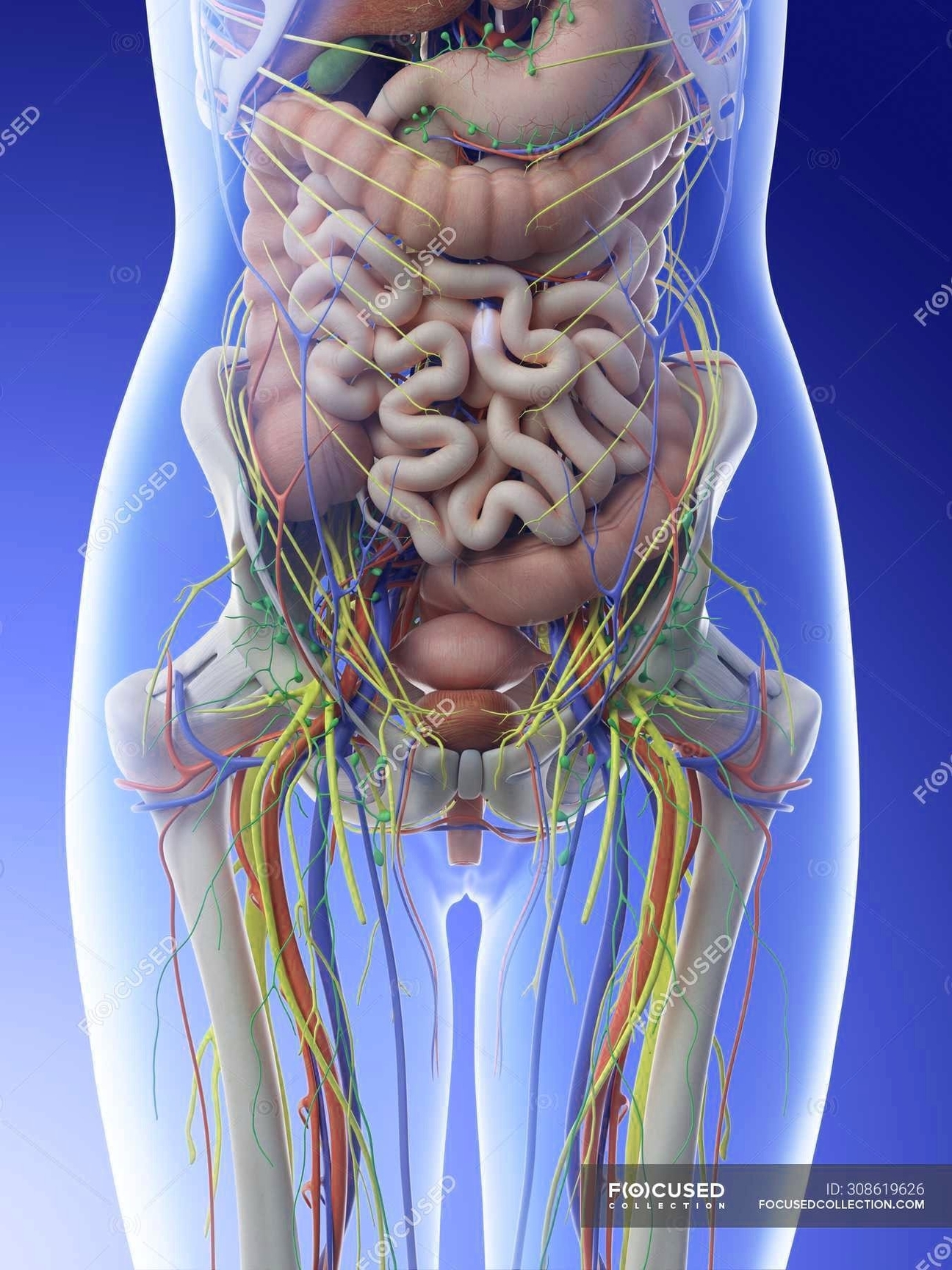
The female abdominal anatomy is a complex and intricate system that houses various organs, each with its unique function. These organs are protected by the abdominal muscles, which include the rectus abdominis in front, the external obliques at the sides, and the latissimus dorsi muscles in the back.
Major Organs
1. Stomach, Small Intestine, and Large Intestine: These organs are responsible for digestion. They turn nutrients into usable energy and help dispose of solid waste.
2. Liver: Located in the upper right-hand part of the abdominal cavity, under the ribs, the liver processes blood, separating waste from nutrients.
3. Gallbladder: This tiny sack under the liver holds extra bile made by the liver until it is pumped into the small intestine. Bile helps break down fat.
4. Pancreas: This gland produces enzymes to help your body digest proteins, carbohydrates, and fats. It also makes hormones that help regulate the distribution of nutrients, including sugar.
5. Kidneys: Located near the back of the body, under the ribs, on each side of the spine, kidneys filter waste out of the bloodstream, which is passed out of the body as urine. They also help regulate levels of electrolytes, like salt and potassium, and produce certain hormones.
6. Suprarenal (Adrenal) Glands: These glands synthesize and secrete hormones that help the kidneys conserve sodium, thus conserving water. They also play a role in supporting the bodys sexual functions.
Female Reproductive Organs
1. Uterus (Womb): A hollow, pear-shaped organ located in a woman’s lower abdomen, between the bladder and the rectum.
2. Ovaries: Two female reproductive organs located in the pelvis.
3. Fallopian Tubes: These carry eggs from the ovaries to the uterus.
4. Cervix: The lower part of the uterus that opens into the vagina.
5. Vagina: The canal that joins the cervix to the outside of the body.
External Female Anatomy
1. Mons Pubis: The rounded, fleshy area on the front of the pelvic bone where pubic hair usually grows.
2. Labia Majora and Minora: The fleshy outer and inner folds of protective skin located on each side of the vaginal opening.
3. Clitoris: Sits at the top of the vulva, above the urethral opening.
4. Urethral Opening: The tube that carries urine from the bladder to the outside of the body.
These organs and structures work together to perform a variety of functions, including digestion, waste removal, reproduction, and hormone regulation. Understanding the female abdominal anatomy is crucial for maintaining health and diagnosing potential health issues..