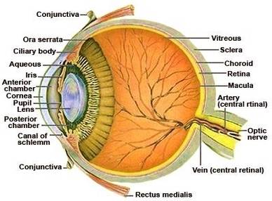
Here are descriptions of some of the main parts of the eye: Cornea: The cornea is the clear outer part of the eye’s focusing system located at the front of the eye. Iris: The iris is the colored part of the eye that regulates the amount of light entering the eye. Lens:
The eye anatomy at the back comprises the following: vitreous humor, retina, choroid and optic nerve. The vitreous humor is the jelly-like substance that fills the vitreous cavity between the lens and the retina. It is transparent and thus allows light to be focused onto the retina.
Complete Physiology of Eye is described below in the given paragraph: The eye is rather like a living Camera. Each eye is a liquid-filled ball 2.5 cm in diameter. At the front of the eye is a clear, round window called the cornea. Behind the cornea is a �lens. A camera focuses by moving the lens nearer or further away from the object.

eye anatomy


