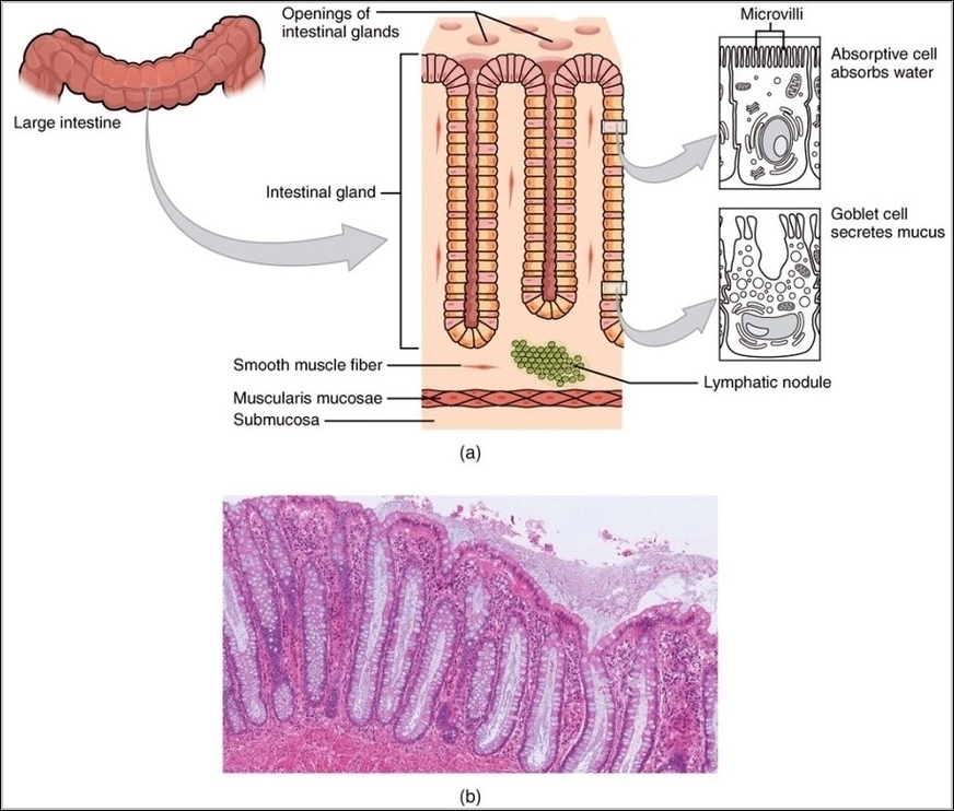
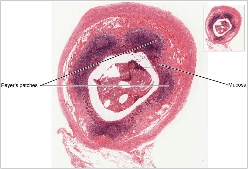
Blood Flow Contracted Ventricles

Blood Flow Contracted Ventricles

A heart cycle image showing contracted ventricles focuses on blood being forced into the aorta and pulmonary artery. Valve positions and flow direction clarify how each heartbeat moves blood efficiently…
Gross Anatomy of the Lungs

Gross Anatomy of the Lungs

The lungs are paired spongy organs in the thoracic cavity, each divided into lobes—three on the right, two on the left—to accommodate the heart's position. The right lung is shorter…
Mucosa Associated Lymphoid Tissue MALT Nodule

Mucosa Associated Lymphoid Tissue MALT Nodule

Mucosa-associated lymphoid tissue, including nodules like Peyer’s patches in ileum, tonsils, and appendix, forms diffuse surveillance networks beneath epithelial surfaces rich in antigens, with M cells sampling lumen contents for…
Internal Anatomy of the HeartN

Internal Anatomy of the Heart

The heart's internal anatomy features four chambers with septa dividing left and right, atria receiving blood via smooth posterior walls and pectinate anterior plus auricles, ventricles with trabeculae carneae and…