5,814 male human anatomy diagram stock photos and images available, or start a new search to explore more stock photos and images. Male silhouette (contour) on white background, vector. Male,…
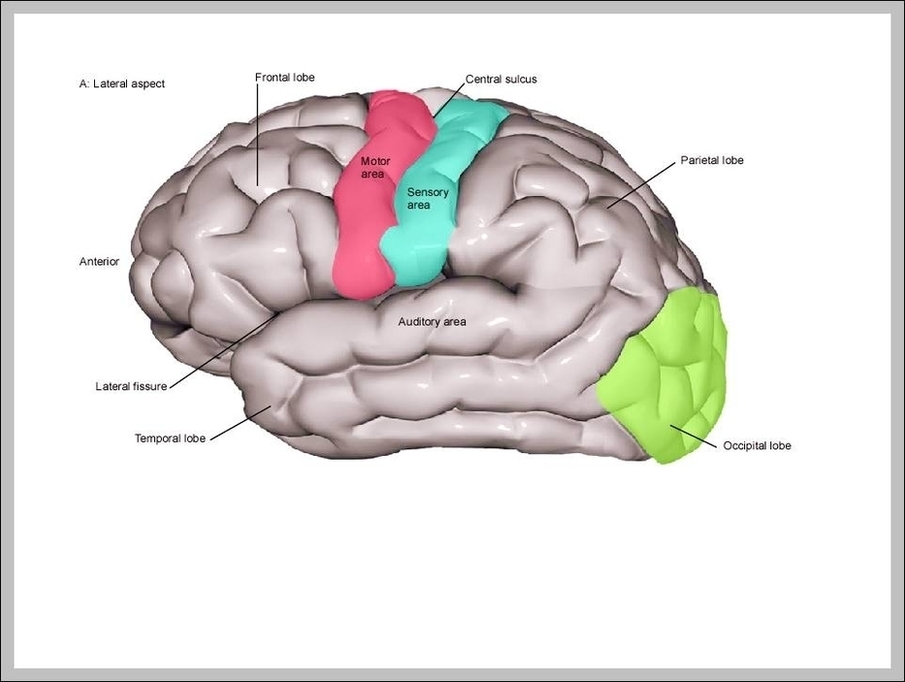
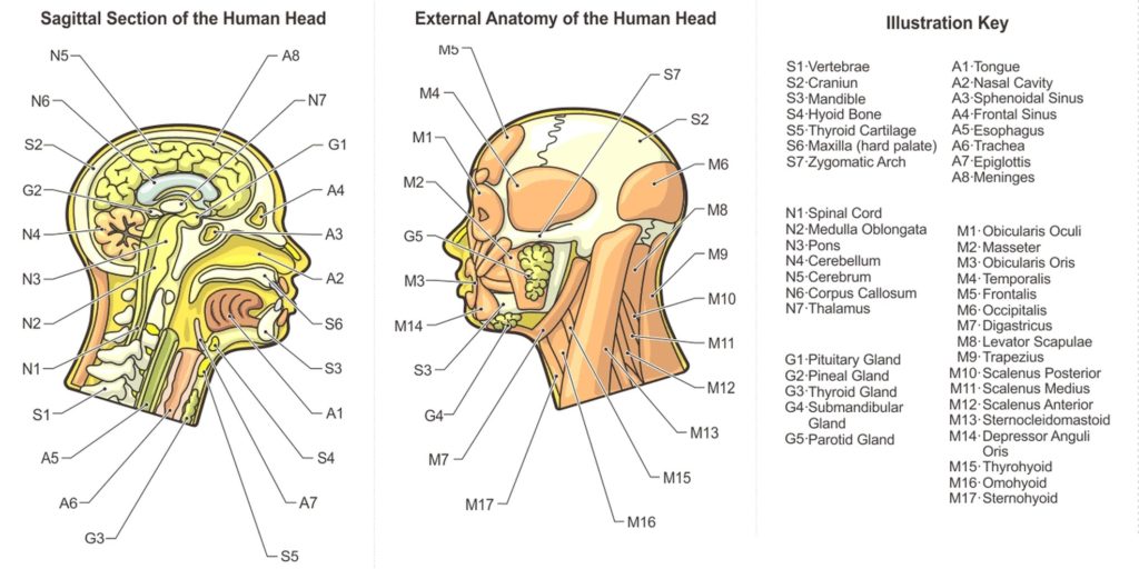
It is a complex anatomical structure weighing up to five kilograms that rests on the bony skull and in turn, the neck. In addition to the evident ears, eyes, nose,…
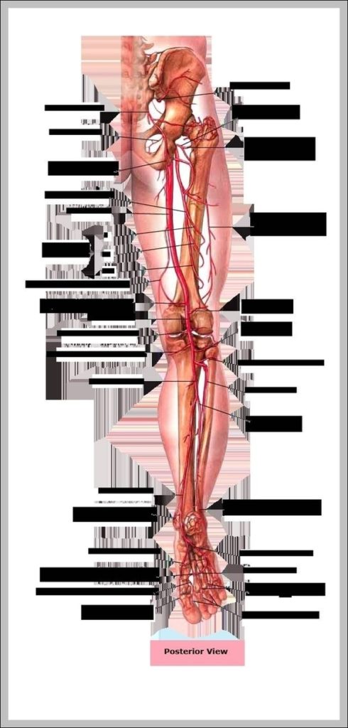
4,236 human leg anatomy stock photos and images available, or start a new search to explore more stock photos and images. The high-quality vector images of the human anatomy are…
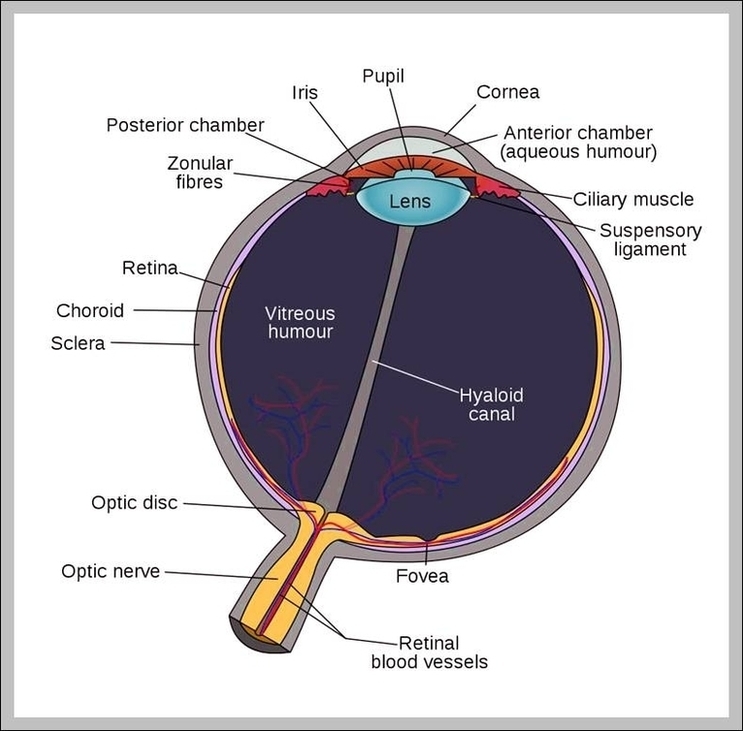
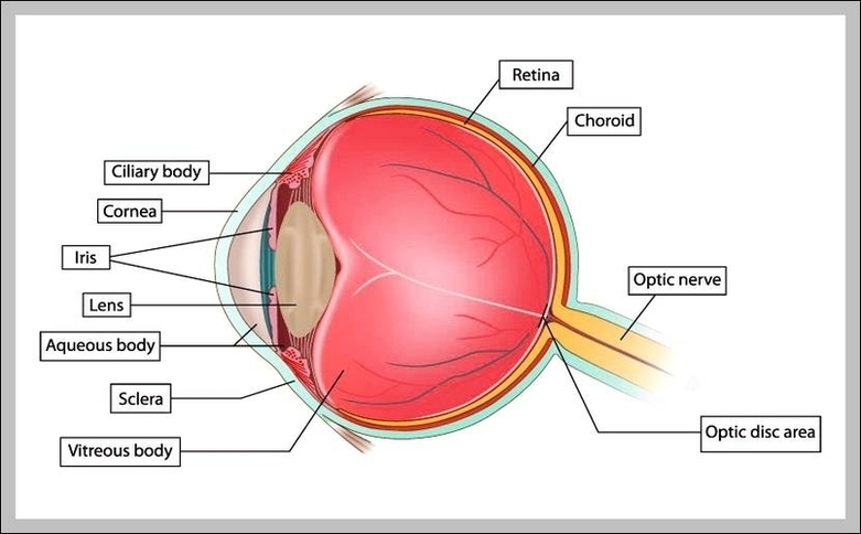
The Human Eye (Eyeball) Diagram, Parts and Pictures. The human eye consists of the eyeball, optic nerve, orbit and appendages (eyelids, extraocular muscles and lacrimal glands). While the eyeball is…
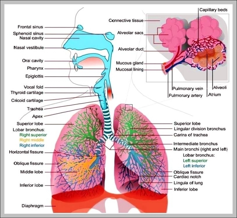
The respiratory system is the network of organs and tissues that help you breathe. It includes your airways, lungs, and blood vessels. The muscles that power your lungs are also…
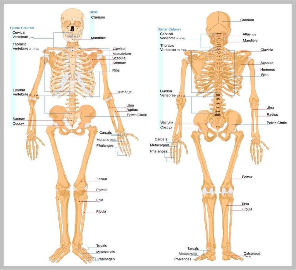
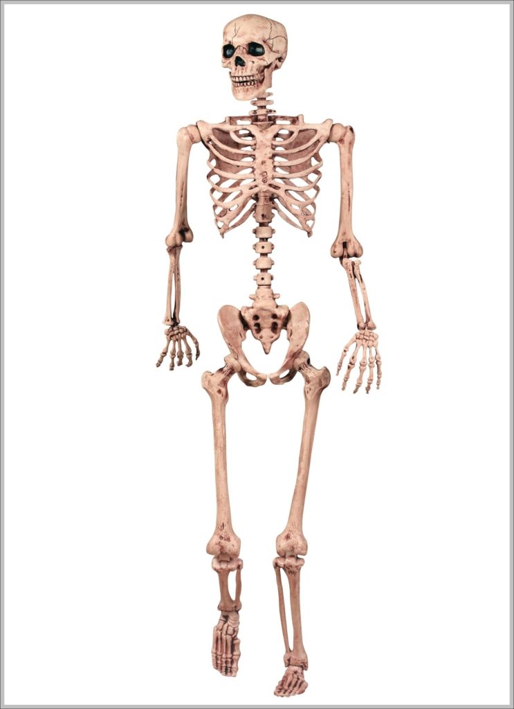
31,815 human skeleton anatomy stock photos and images available, or search for human bones or human anatomy to find more great stock photos and pictures. Human skeleton, the internal skeleton…
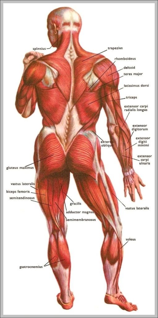
Muscle Cell Types 1 Skeletal Muscle. Skeletal muscle is the most common and widely distributed muscle tissue in the body, making up around 40% of the body’s total mass. 2…
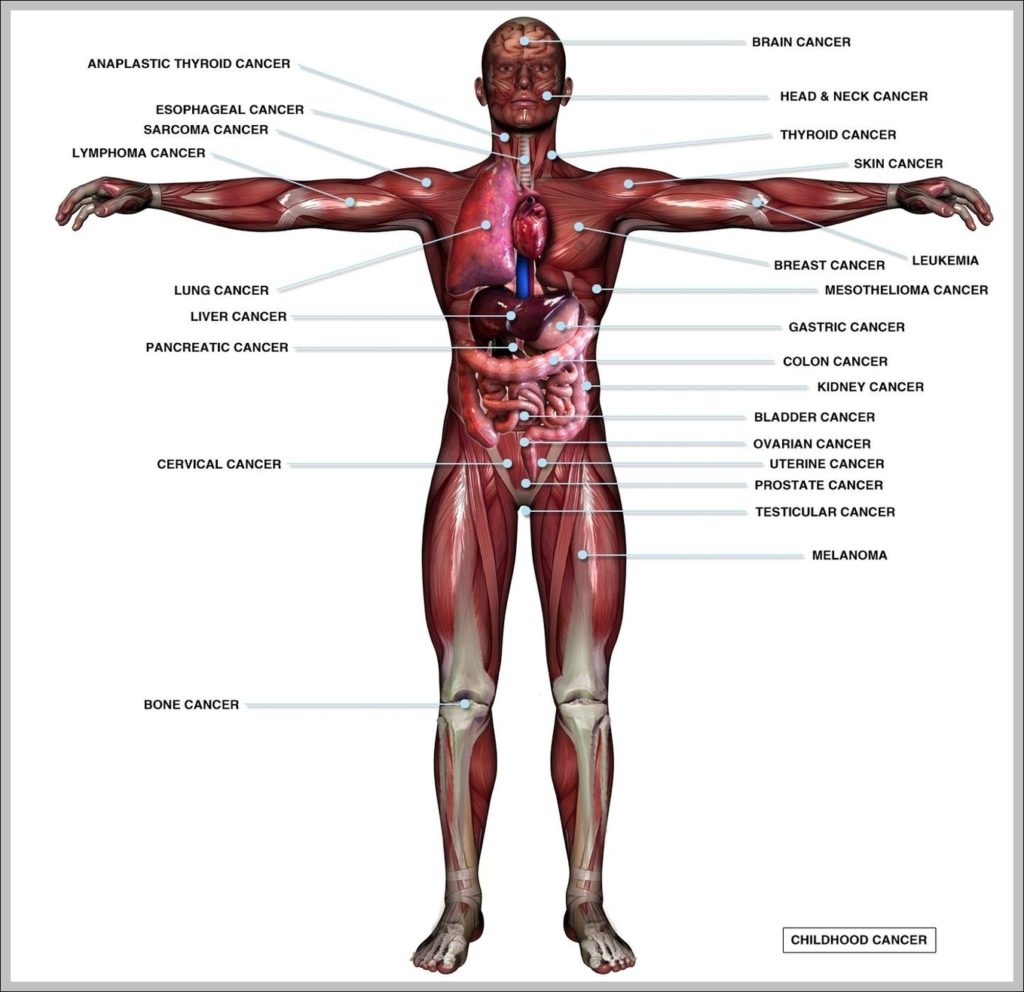
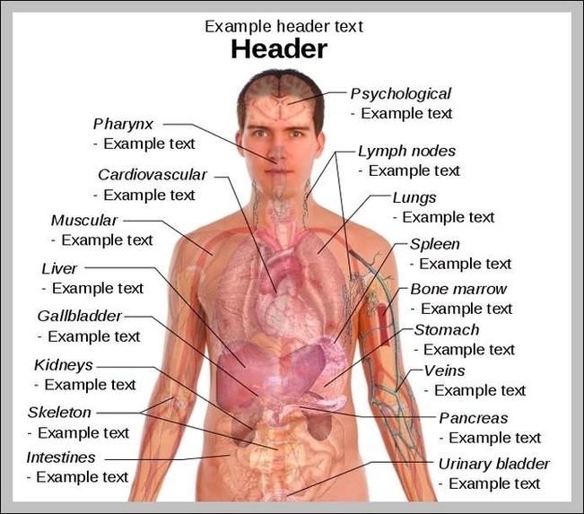
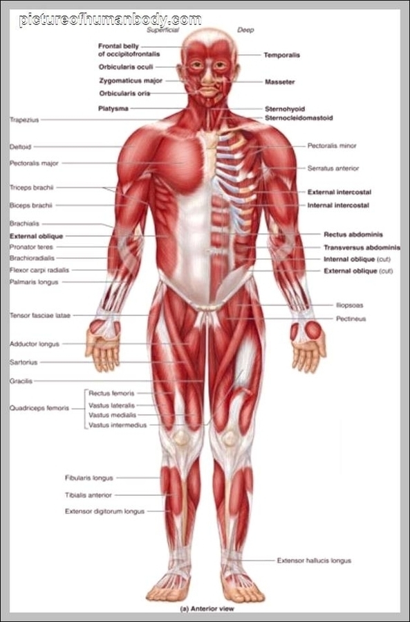
9,591 human torso anatomy stock photos and images available, or search for human anatomy to find more great stock photos and pictures. Male Torso stock illustrations A torso of human…
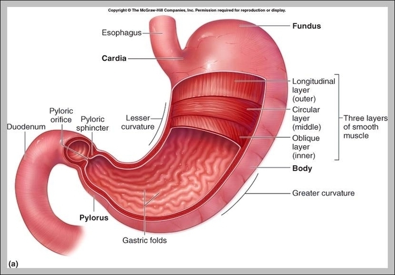
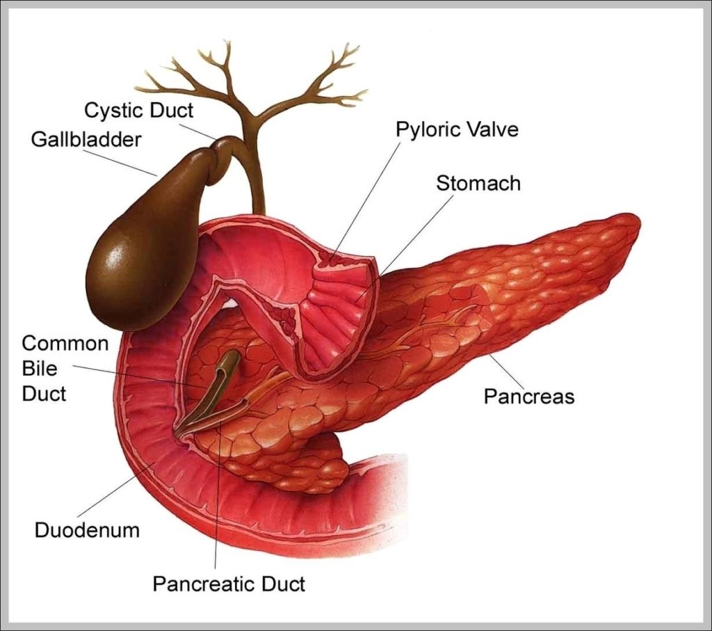
2,755 human stomach anatomy stock photos and images available or start a new search to explore more stock photos and images. Next. The stomach is a muscular organ located on…
Common ways to learn anatomy online include YouTube videos and online multimedia learning platforms such as Kenhub. There are several fantastic YouTube channels available for learning anatomy. For those who…
Abstract image human body in the form of a starry sky or space, consisting of points, lines, and shapes in the form of planets, stars and the universe. Low poly…
A hinge joint is a common class of synovial joint that includes the ankle, elbow, and knee joints. Hinge joints are formed between two or more bones where the bones…
45,936 human skeleton stock photos and images available, or search for human skeleton anatomy or human skeleton vector to find more great stock photos and pictures. Skeleton in various poses…
Taste bud and the papillae of the tongue. basic taste areas: sweet, salty, sour, bitter and umami. Taste bud and the papillae of the tongue. Human mouth isolated Taste bud.…
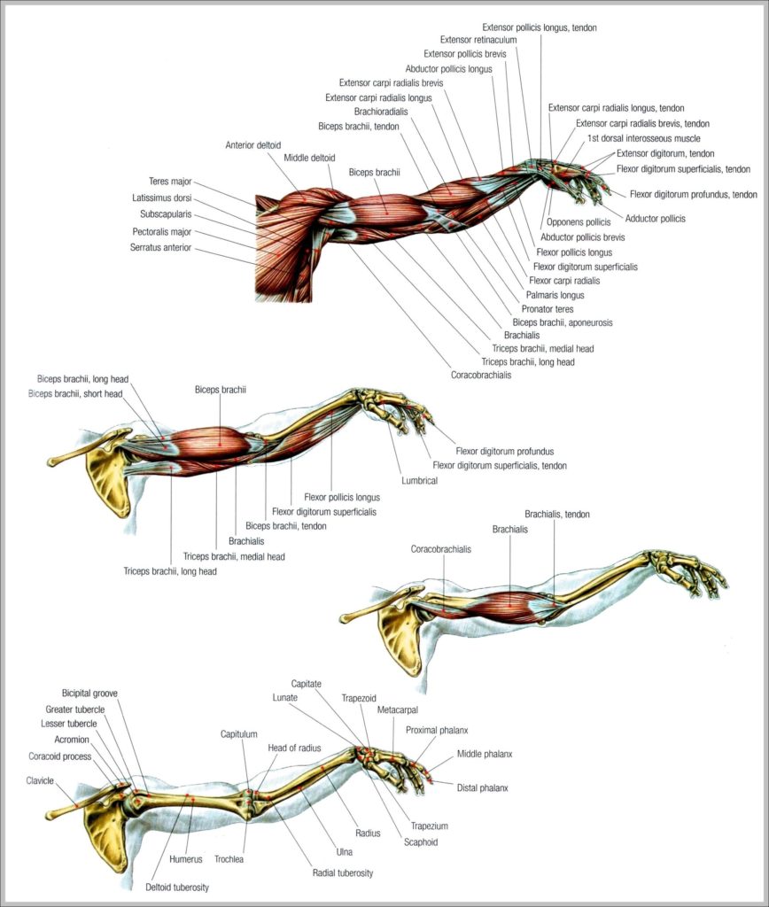
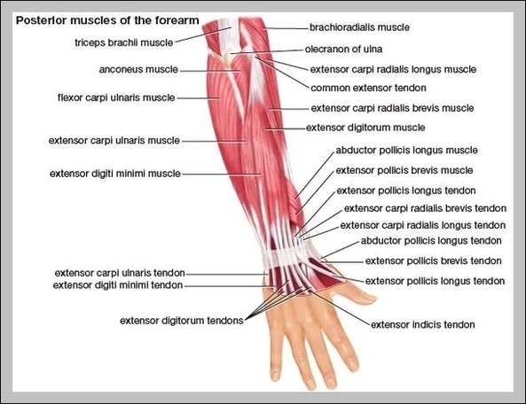
The muscles of the forearm can be divided into two groups: anterior (flexors) and posterior (extensors). Both the flexors and extensors are further divided into superficial and deep layers. The…
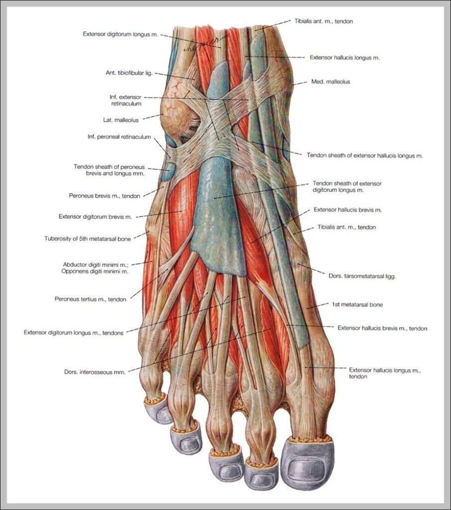
The muscles of the foot can be split into two groups, the extrinsic and intrinsic muscles. The extrinsic foot muscles are found in the lower leg and act to dorsiflex,…