anterior torso muscles. These intricate structures play a crucial role in our daily activities, from breathing to movement. Buckle up as we explore their anatomy and functions.
## Anterior Trunk Muscles: Guardians of the Front
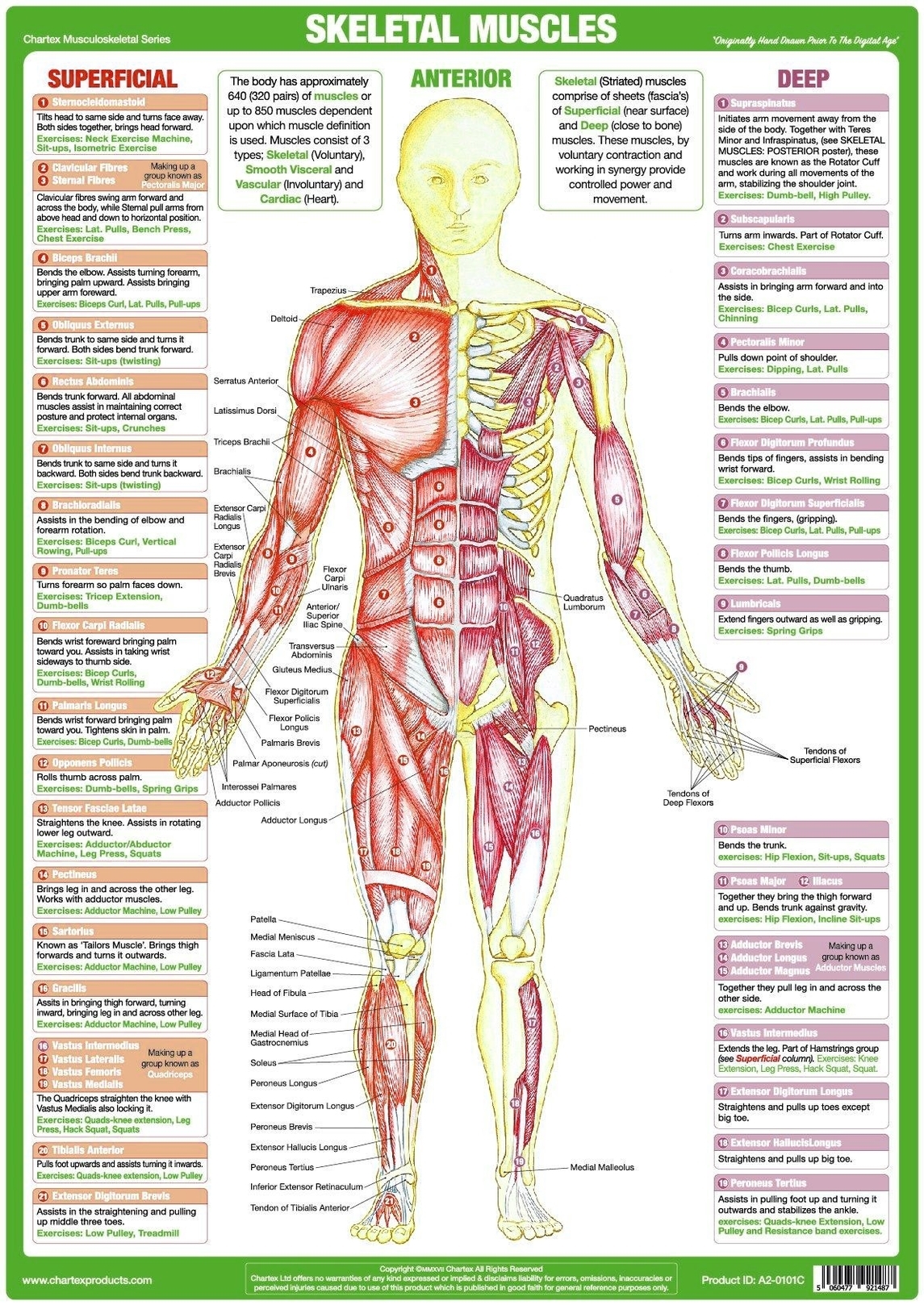
The anterior trunk muscles reside on the front and sides of the torso, attaching to the bony framework of the thoracic cage and pelvis. They form a robust network that supports and moves the upper body. Let’s break down these muscles into key groups:
### 1. Muscles of the Thoracic Cage
#### a. Pectoralis Major:
– The pectoralis major is a powerful muscle that spans the chest. It originates from the clavicle, sternum, and upper ribs, and inserts onto the humerus.
– Its primary functions include flexing and adducting the arm, making it essential for movements like hugging, pushing, and lifting.
#### b. Pectoralis Minor:
– The pectoralis minor lies beneath the pectoralis major. It originates from the third to fifth ribs and attaches to the scapula.
– This muscle assists in stabilizing the scapula during arm movements.
#### c. Serratus Anterior:
– The serratus anterior runs along the lateral rib cage. It originates from the upper ribs and inserts onto the scapula.
– Its unique arrangement resembles serrated teeth, hence the name. It plays a crucial role in protracting and rotating the scapula, aiding movements like reaching forward and lifting objects.
#### d. Subclavius:
– The subclavius muscle lies beneath the clavicle. It connects the clavicle to the first rib.
– Although small, it stabilizes the clavicle and assists in depressing the shoulder.
#### e. Intercostal Muscles:
– The intercostal muscles are found between adjacent ribs. They come in three layers: external, internal, and innermost.
– These muscles facilitate rib movement during breathing. The external intercostals elevate the ribs during inhalation, while the internal and innermost intercostals aid exhalation.
#### f. Subcostals:
– The subcostals are deep muscles that run parallel to the ribs. They assist in rib depression during exhalation.
#### g. Transversus Thoracis:
– The transversus thoracis lies deep within the chest. It originates from the sternum and inserts onto the ribs.
– This muscle contributes to rib stabilization and assists in exhalation.
#### h. Diaphragm:
– The diaphragm is a large, dome-shaped muscle that separates the thoracic and abdominal cavities.
– It plays a pivotal role in breathing, contracting during inhalation and relaxing during exhalation.
### 2. Anterolateral Abdominal Wall Muscles
#### a. Rectus Abdominis:
– The rectus abdominis, commonly known as the “six-pack,” runs vertically along the midline of the abdomen.
– It flexes the trunk, aids in sitting up, and provides stability during movements.
#### b. External Abdominal Oblique:
– The external abdominal oblique forms the outermost layer of the abdominal wall.
– It assists in trunk rotation, lateral flexion, and compressing the abdomen.
#### c. Internal Abdominal Oblique:
– The internal abdominal oblique lies beneath the external oblique.
– It performs similar functions, including trunk rotation and lateral flexion.
#### d. Transversus Abdominis:
– The transversus abdominis is the deepest abdominal muscle. It runs horizontally across the abdomen.
– Its primary role is abdominal compression, crucial for maintaining posture and supporting the spine.
#### e. Pyramidalis:
– The pyramidalis is a small triangular muscle located just above the pubic bone.
– While its function isn’t fully understood, it likely contributes to tension in the linea alba (a fibrous band in the midline of the abdomen).
#### f. Quadratus Lumborum:
– The quadratus lumborum lies deep in the lower back. It connects the pelvis to the lower ribs.
– This muscle assists in lateral flexion of the trunk and helps maintain an upright posture.
## Conclusion
These anterior torso muscles are the unsung heroes of our daily movements. Whether we’re lifting groceries, taking a deep breath, or simply sitting up, they work tirelessly to keep us going. So next time you stretch or twist, remember