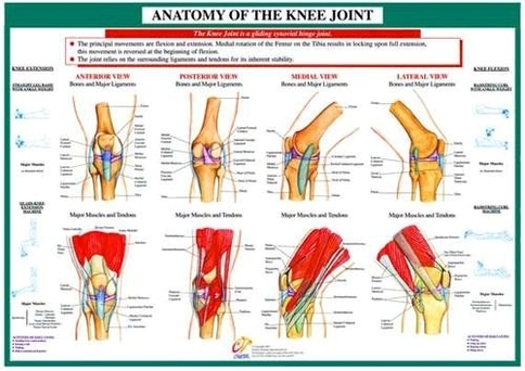
The shin bone (tibia), the thigh bone (femur), and the kneecap (patella) are each important parts of the knee joint. A fourth bone, the fibula, is located just next to the shin bone (tibia) and knee joint, and can play an important role in some knee conditions. The tibia, femur, and patella,…
Bones Around the Knee. A fourth bone, the fibula, is located just next to the shin bone (tibia) and knee joint, and can play an important role in some knee conditions. The tibia, femur, and patella, all are covered with a smooth layer of cartilage (see below) where they contact each other at the knee joint.
Bones Around the Knee. A fourth bone, the fibula, is located just next to the shin bone (tibia) and knee joint, and can play an important role in some knee conditions. The tibia, femur, and patella, all are covered with a smooth layer of cartilage (see below) where they contact each other at the knee joint.

Anatomy of the knee joint
Posted inDiagrams


