The stomach, a vital organ in the human digestive system, plays a central role in the processing of food. Let’s delve into its intricate anatomy, function, and significance.
## Anatomy of the Stomach
1. Location:
– The stomach resides in the upper abdomen, nestled between the esophagus (the tube connecting the throat to the stomach) and the duodenum (the initial part of the small intestine).
– Its J-shaped structure allows it to accommodate varying food volumes.
2. Coverings and Connections:
– The stomach is enveloped by a layer of peritoneum, a serous membrane that also connects it to neighboring organs.
– The lesser omentum links the stomach to the liver and then wraps around it.
– The greater omentum, akin to a curtain, extends downward from the stomach.
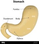
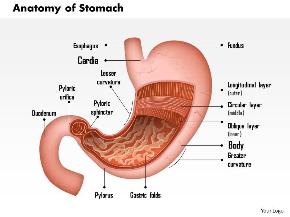
3. Parts of the Stomach:
– Cardia: The uppermost part, near the esophagus.
– Fundus: The dome-shaped region above the cardia.
– Body: The main central portion.
– Pyloric part: The lower section leading to the duodenum.
4. Function of the Stomach:
– Accumulation: The stomach stores ingested food, allowing for controlled release into the small intestine.
– Digestion: It initiates both mechanical (churning) and chemical (acid and enzymes) digestion.
– Hydrochloric Acid: The stomach secretes potent hydrochloric acid, essential for breaking down proteins and activating enzymes.
– Gastric Juices: These juices contain enzymes like pepsin, which further digest proteins.
5. Layers of the Stomach Wall:
– Mucosa: The innermost layer, featuring gastric pits and glands. It secretes mucus, acid, and enzymes.
– Submucosa: Contains blood vessels, nerves, and connective tissue.
– Muscularis Externa: Comprises three muscle layers (longitudinal, circular, and oblique) for peristalsis.
– Serosa: The outermost layer, covered by peritoneum.
6. Blood Supply:
– Gastric Arteries: Supply blood to the stomach.
– Gastroomental Arteries: Branches that nourish specific regions.
– Short Gastric Arteries and Posterior Gastric Arteries: Additional blood supply.
– Gastroduodenal Artery: Feeds both the stomach and duodenum.
7. Innervation:
– Parasympathetic: The vagus nerve (Cranial Nerve X) regulates stomach activity.
– Sympathetic: The celiac plexus (T5-T12) provides sympathetic innervation.
8. Lymphatics:
– Lymph nodes associated with the stomach include the juxtacardial, gastric, short gastric, gastroomental, and pyloric nodes.
– These nodes eventually drain into the celiac nodes, forming part of the intestinal lymphatic system.
9. Clinical Point:
– Hiatal Hernia: A condition where part of the stomach protrudes through the diaphragm into the chest cavity.
In summary, the stomach’s intricate design enables it to process food efficiently, making it a crucial player in our daily lives. Remember, while it may not be able to pick up new hobbies, it certainly wields the power to corrode metalthanks to its formidable hydrochloric acid! ?????
For a deeper exploration, consider studying the peritoneal course and the entire digestive system, tracing the path of this remarkable organ within our abdominal cavity..
o