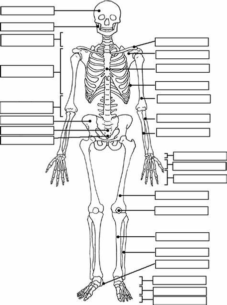
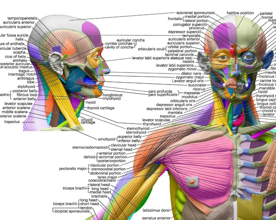
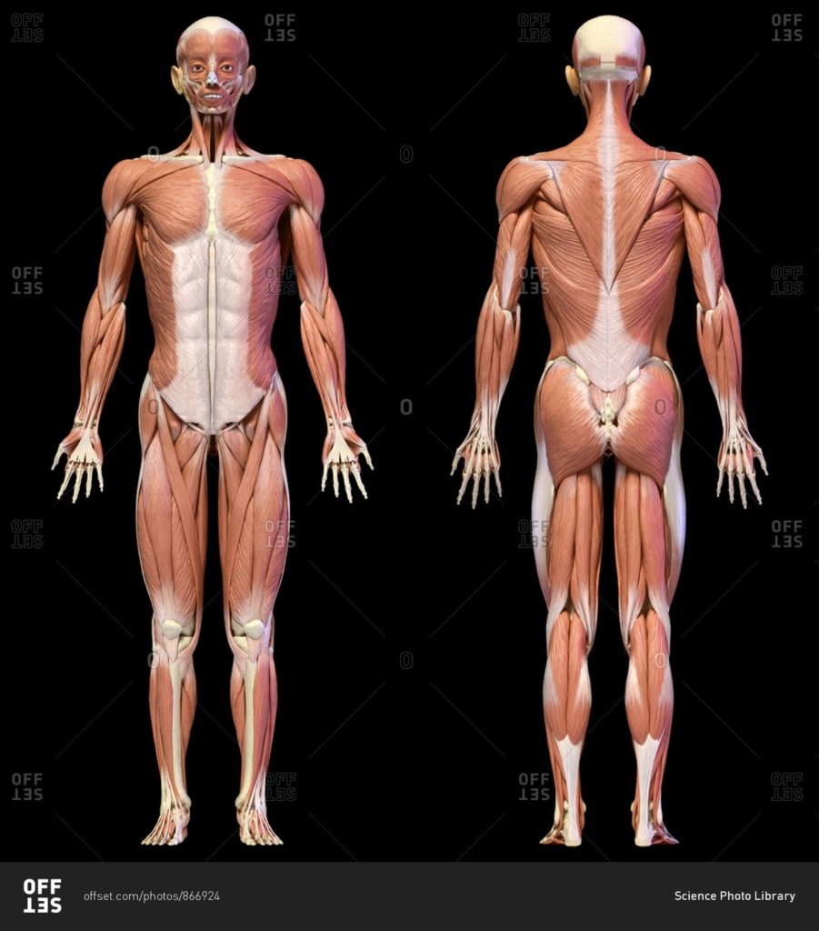
Human Anatomy Skeletal System

Human Anatomy: The Skeletal System

The skeletal system, also known as the musculoskeletal system, is a vital part of the human body. It provides the body with shape, stability, and protection of internal organs. It is composed of 206 bones that connect to each other via joints. Accessory structures that support the skeletal system are the cartilage, ligaments, bursae, and muscle tendons.

The skeletal system can be broken down into two parts: the axial skeleton and the appendicular skeleton.

Axial Skeleton

The adult axial skeleton consists of 80 bones. It’s made up of the bones that form the vertical axis of the body, such as the bones of the head, neck, chest, and spine.

*Skull Bones*: The adult skull comprises 22 bones. These bones can be further classified by location: Cranial bones (8) form the bulk of your skull and help to protect your brain. Facial bones (14) are found on the front of the skull and make up the face.

*Auditory Ossicles*: The auditory ossicles are six small bones found within the inner ear canal in the skull. They work together to transmit sound waves from the surrounding environment to the structures of the inner ear.

*Hyoid*: The hyoid is a U-shaped bone found at the base of the jaw. It serves as a point of attachment for muscles and ligaments in the neck.

*Vertebral Column*: The vertebral column is made up of 26 bones. The first 24 are all vertebrae, followed by the sacrum and coccyx (tailbone).

*Thoracic Cage*: The thoracic cage is made up of the sternum (breastbone) and 12 pairs of ribs. These bones form a protective cage around the organs of the upper torso, including the heart and lungs.

Appendicular Skeleton

There are a total of 126 bones in the appendicular skeleton. It consists of the bones that make up the arms and legs, as well as the bones that attach them to the axial skeleton.

The skeletal system is more than just a framework. It also plays a crucial role in movement, blood cell production, mineral storage, and organ protection. Despite its rigid structure, the skeletal system is dynamic, constantly remodeling and adapting to physical stress.

The skeletal system is also unique in that it undergoes significant changes throughout a person’s life. Children’s skeletons actually contain more bones because some of them, including those of the skull, fuse together as they grow up. There are also some differences in the male and female skeleton. The male skeleton is usually longer and has a high bone mass. The female skeleton, on the other hand, has a broader pelvis to accommodate for pregnancy and childbirth.

In conclusion, the skeletal system is a complex and vital component of human anatomy. Its intricate design and multifaceted functions underscore its importance in supporting life and health.

Human Anatomy Skeletal System Diagram - Human Anatomy Skeletal System Chart - Human anatomy diagrams and charts explained. This anatomy system diagram depicts Human Anatomy Skeletal System with parts and labels. Best diagram to help learn about health, human body and medicine.

Human Anatomy Skeletal System

Organ System Biology

Organ System Biology

Organ System Biology is a branch of biology that studies the structure and function of various organ systems in the body. An organ system is a group of organs that work together to perform a specific function or set of functions in an organism’s body. The human body is made up of multiple organ systems, each with a specialized role.

Levels of Organization

The body has levels of organization that build on each other. Cells make up tissues, tissues make up organs, and organs make up organ systems. Each organ has a specialized role in a plant or animal body, and is made up of distinct tissues.

Key Organ Systems

1. Integumentary System: Consists of external organs that protect the body from damage, including the skin, fingernails, and hair.

2. Skeletal System: Made up of all the bones in the human body, forming the supporting structure of the body.

3. Muscular System: Includes the different types of muscles in the body: cardiac, smooth, and skeletal muscles.

4. Circulatory System: Consists of the heart, veins, arteries, and capillaries. It circulates blood throughout the body to transport nutrients and oxygen to the cells.

Interdependence of Organ Systems

The function of an organ system depends on the integrated activity of its organs. For instance, digestive system organs cooperate to process food. The survival of the organism depends on the integrated activity of all the organ systems, often coordinated by the endocrine and nervous systems.
pecialized Systems

Most cells in large multicellular organisms don’t directly exchange substances like nutrients and wastes with the external environment. Instead, they are surrounded by an internal environment of extracellular fluid. Humans and other complex organisms have specialized systems that maintain the internal environment, keeping it steady and able to provide for the needs of the cells.

Conclusion

Organ System Biology provides a comprehensive understanding of how the body’s various systems work together to maintain homeostasis and ensure the survival of the organism. It underscores the complexity and interdependence of biological systems, highlighting the intricate balance that sustains life..

Organ System Biology Diagram - Organ System Biology Chart - Human anatomy diagrams and charts explained. This anatomy system diagram depicts Organ System Biology with parts and labels. Best diagram to help learn about health, human body and medicine.

Organ System Biology

Anatomy And Physiology Circulatory System Representation Explained

The circulatory system, also known as the cardiovascular system, is a remarkable network that ensures the delivery of essential substances to every cell in our body. It plays a pivotal role in maintaining homeostasis and sustaining life. Let’s delve into the intricacies of this intricate system.

## Anatomy of the Circulatory System

### 1. The Heart
At the core of this system lies the heart, a muscular organ that tirelessly pumps blood throughout our body. It has four chambers: two atria (upper chambers) and two ventricles (lower chambers). The right side of the heart receives oxygen-poor blood from the body and pumps it to the lungs for oxygenation. The left side receives oxygen-rich blood from the lungs and propels it to the rest of the body via a vast network of blood vessels.

### 2. Blood Vessels
The circulatory system comprises three types of blood vessels:

– Arteries: These sturdy vessels carry oxygenated blood away from the heart. The largest artery, the aorta, branches into smaller arteries that reach every tissue and organ.

– Veins: Veins transport deoxygenated blood back to the heart. The superior and inferior vena cava collect blood from various body parts and deliver it to the right atrium.

– Capillaries: These microscopic vessels connect arteries and veins. Capillaries facilitate the exchange of gases, nutrients, and waste products between blood and surrounding tissues.

### 3. Blood
Our blood is a dynamic fluid composed of several components:

– Red Blood Cells (Erythrocytes): These disc-shaped cells contain hemoglobin, which binds to oxygen. Erythrocytes transport oxygen from the lungs to body tissues.

– White Blood Cells (Leukocytes): These immune system warriors defend against infections and foreign invaders.

– Platelets (Thrombocytes): These tiny cells play a crucial role in blood clotting, preventing excessive bleeding.

### Circulatory Pathways

1. Pulmonary Circulation:
– Oxygen-poor blood returns to the right atrium via the superior and inferior vena cava.
– The right atrium contracts, pushing blood into the right ventricle.
– The right ventricle pumps blood into the pulmonary artery, which splits into vessels leading to both lungs.
– In the lungs, blood picks up oxygen and releases carbon dioxide.
– Oxygenated blood returns to the left atrium via the pulmonary veins.

2. Systemic Circulation:
– From the left atrium, blood enters the left ventricle.
– The left ventricle propels oxygen-rich blood into the aorta.
– The aorta branches into smaller arteries, which supply blood to all body tissues.
– Capillaries facilitate nutrient and gas exchange.
– Deoxygenated blood returns to the heart via veins, ultimately reaching the superior and inferior vena cava.

3. Coronary Circulation:
– The heart itself needs nourishment. Coronary arteries branch off the aorta and supply blood to the heart muscle.
– The right coronary artery and the left coronary artery ensure adequate blood flow to the heart.

### Clinical Considerations

– Arteriosclerosis: Thickening and hardening of arterial walls.
– Cerebrovascular Disease: Disorders affecting blood vessels in the brain.
– Peripheral Artery Disease: Reduced blood flow to limbs.
– Aneurysm: Abnormal dilation of an artery.
– Varices: Enlarged veins, often seen in the legs.
– Arrhythmia: Irregular heart rhythm.
– Heart Failure: Inability of the heart to pump effectively.

In summary, the circulatory system orchestrates an intricate dance of blood, oxygen, and nutrients, ensuring our survival. Its ceaseless rhythm echoes life itself, silently sustaining us day after day..

Anatomy And Physiology Circulatory System Representation Explained Diagram - Anatomy And Physiology Circulatory System Representation Explained Chart - Human anatomy diagrams and charts explained. This anatomy system diagram depicts Anatomy And Physiology Circulatory System Representation Explained with parts and labels. Best diagram to help learn about health, human body and medicine.

Anatomy And Physiology Circulatory System Representation Explained

Overview Of The Digestive System

Overview of the Digestive System

The digestive system is a complex network of organs that work together to break down food, absorb nutrients, and eliminate waste. It is composed of the gastrointestinal (GI) tract, also known as the digestive tract, and the liver, pancreas, and gallbladder.

The GI tract is a series of hollow organs joined in a long, twisting tube from the mouth to the anus. The hollow organs that make up the GI tract are the mouth, esophagus, stomach, small intestine, large intestine, and anus. The liver, pancreas, and gallbladder are the solid organs of the digestive system.

The small intestine has three parts: the duodenum, jejunum, and ileum. The large intestine includes the appendix, cecum, colon, and rectum. The appendix is a finger-shaped pouch attached to the cecum. The cecum is the first part of the large intestine. The colon is next. The rectum is the end of the large intestine.

Bacteria in your GI tract, also called gut flora or microbiome, help with digestion. Parts of your nervous and circulatory systems also help. Working together, nerves, hormones, bacteria, blood, and the organs of your digestive system digest the foods and liquids you eat or drink each day.

Digestion is important because your body needs nutrients from food and drink to work properly and stay healthy. Proteins, fats, carbohydrates, vitamins, minerals, and water are nutrients. Your digestive system breaks nutrients into parts small enough for your body to absorb and use for energy, growth, and cell repair.

Each part of your digestive system helps to move food and liquid through your GI tract, break food and liquid into smaller parts, or both. Once foods are broken into small enough parts, your body can absorb and move the nutrients to where they are needed. Your large intestine absorbs water, and the waste products of digestion become stool. Nerves and hormones help control the digestive process.

In conclusion, the digestive system is a vital part of the human body. It is responsible for breaking down food, absorbing nutrients, and eliminating waste. Without it, our bodies would not be able to function properly. It is a complex system that requires all of its parts to work together in order to function efficiently.

Overview Of The Digestive System Diagram - Overview Of The Digestive System Chart - Human anatomy diagrams and charts explained. This anatomy system diagram depicts Overview Of The Digestive System with parts and labels. Best diagram to help learn about health, human body and medicine.

Overview Of The Digestive System

Female Reproductive System Illustrated

The female reproductive system is a complex and intricate structure that plays a crucial role in sexual pleasure, reproduction, and overall health. It consists of both internal and external structures.

External Female Anatomy

The external structures, collectively referred to as the vulva, include:

1. Mons Pubis: The rounded, fleshy area on the front of the pelvic bone where pubic hair usually grows.
2. Labia Majora: The fleshy outer folds of protective skin located on each side of the vaginal opening.
3. Labia Minora: The skinfolds that are just inside the labia majora.
4. Clitoris: Located at the top of the vulva, above the urethral opening. A fold of skin called the clitoral hood covers most of the clitoris, leaving only the tip visible.
5. Urethral Opening: The tube that carries urine from the bladder to the outside of the body.

Internal Female Anatomy

The internal structures include:

1. Vagina: A muscular canal that extends from the cervix, which is the lower part of the uterus, to the external part of the female genitalia, which is the vulva.
2. Cervix: The lower part of the uterus that connects to the vagina.
3. Uterus: Also known as the womb, the uterus is a hollow, pear-shaped organ where a fetus grows.
4. Ovaries: Two oval-shaped organs that produce, store, and release eggs into the fallopian tubes in the process known as ovulation.
5. Fallopian Tubes: These tubes transport the eggs from the ovaries to the uterus.
6. Hymen: A thin membrane that partially covers the vaginal opening.
7. Bartholin Glands and Skene’s Glands: These glands provide lubrication during sexual intercourse.

Breasts

Female breasts have both internal and external parts. They play a significant role in breastfeeding.

Functions of the Female Reproductive System

The female reproductive system plays a vital role in sexual pleasure, getting pregnant, and breastfeeding. The urinary system, closely linked with the reproductive system, helps rid the body of toxins through urination.

Illustrations

Illustrations of the female reproductive system provide a visual understanding of the system’s structure and function. They can be found in various medical textbooks, online resources, and educational platforms.

In conclusion, the female reproductive system is a marvel of nature, designed to carry out the miracle of life. Its complexity and functionality are a testament to the intricate design of the human body..

Female Reproductive System Illustrated Diagram - Female Reproductive System Illustrated Chart - Human anatomy diagrams and charts explained. This anatomy system diagram depicts Female Reproductive System Illustrated with parts and labels. Best diagram to help learn about health, human body and medicine.

Female Reproductive System Illustrated

Human Body Skeletal System

The Human Body Skeletal System

The human skeletal system is a complex structure that serves as the body’s support framework. It consists of bones, cartilage, tendons, and ligaments, all of which work together to provide support, protection, and movement.

Composition

The human skeletal system consists of 206 bones in adults. Children’s skeletons contain more bones, which fuse together as they grow up. The skeletal system can be divided into two main parts: the axial skeleton and the appendicular skeleton.

1. Axial Skeleton: The axial skeleton consists of 80 bones, forming the vertical axis of the body. It includes the bones of the head, neck, chest, and spine. The skull comprises 22 bones, further classified into cranial bones (8) and facial bones (14). The vertebral column is made up of 26 bones, including the cervical vertebrae (7), thoracic vertebrae (12), lumbar vertebrae (5), sacrum, and coccyx. The thoracic cage, composed of the sternum and 12 pairs of ribs, protects the organs of the upper torso.

2. Appendicular Skeleton: The appendicular skeleton consists of 126 bones. It includes the bones that make up the arms and legs, as well as the bones that attach them to the axial skeleton.

Functions

The skeletal system performs several crucial functions:

1. Support: The skeletal system provides the structural framework for the human body, giving it shape. The vertebral column, corresponding to the notochord in lower organisms, is the main support of the trunk.

2. Protection: The skeletal system protects our vital organs. The brain is well protected by the cranium, and the spinal cord by the vertebral column. The thoracic cage protects the heart and lungs.

3. Movement: The skeletal system, in conjunction with the muscular system, facilitates body movement.

4. Blood Cell Production: The skeletal system is responsible for the production of blood cells.

5. Mineral Storage: The skeletal system stores minerals, particularly calcium and phosphorus.

Differences in Male and Female Skeletons

The male skeleton is usually longer and has a higher bone mass, while the female skeleton has a broader pelvis to accommodate for pregnancy and childbirth.

Conclusion

The human skeletal system is a marvel of biological engineering. Its intricate design and multifunctionality enable us to perform a wide range of activities, from simple movements to complex athletic feats. Understanding its structure and functions can provide valuable insights into human health and physiology.

Human Body Skeletal System Diagram - Human Body Skeletal System Chart - Human anatomy diagrams and charts explained. This anatomy system diagram depicts Human Body Skeletal System with parts and labels. Best diagram to help learn about health, human body and medicine.

Human Body Skeletal System

Human Respiratory System Visual

The Human Respiratory System

The human respiratory system is a complex network of organs and structures responsible for the vital process of respiration, which involves the exchange of gases between the air we breathe and our body’s cells.

Upper Respiratory System

The upper respiratory system comprises the nose, nasal cavities, and the paranasal sinuses. These structures provide airways for respiration. The pharynx, also part of the upper respiratory system, connects the nasal and oral cavities to the larynx and esophagus. The larynx and vocal cords allow us to breathe, talk, and sing.

Lower Respiratory System

The lower respiratory system consists of the larynx, trachea, bronchi, and the structures within the lungs, such as the bronchioles and alveolar ducts. The trachea is the main airway to the lungs, while the bronchi are passageways that facilitate the movement of air in and out of the lungs.

Lungs and Alveoli

The lungs, located in the thorax and protected by the bony and muscular thoracic cage, are responsible for gas exchange between the air we breathe and our bodies. The alveoli, microscopic air sacs within the lungs, are the site of this external respiration.

Diaphragm

The diaphragm, the main respiratory muscle, plays an essential role in the physical process of breathing. Under the control of the central nervous system, the diaphragm and the intercostal muscles of the chest wall generate a pumping action on the lung, expanding and contracting the internal space of the thorax.

Function of the Respiratory System

Through breathing, inhalation and exhalation, the respiratory system facilitates the exchange of gases between the air and the blood and between the blood and the body’s cells. The respiratory system also helps us to smell and create sound.

Conclusion

The human respiratory system is a marvel of biological engineering, efficiently facilitating the exchange of life-sustaining gases. Understanding its structure and function is crucial to comprehending how our bodies utilize oxygen and expel waste gases, maintaining the delicate balance necessary for life..

Human Respiratory System Visual Diagram - Human Respiratory System Visual Chart - Human anatomy diagrams and charts explained. This anatomy system diagram depicts Human Respiratory System Visual with parts and labels. Best diagram to help learn about health, human body and medicine.

Human Respiratory System Visual

Human Skeletal System Illustration

The Human Skeletal System

The human skeletal system, also known as the musculoskeletal system, is the internal framework of the body, providing support, enabling movement, and performing several other essential functions.

Composition

The human skeleton is composed of around 270 bones at birth, which decreases to around 206 bones by adulthood after some bones get fused together. The bone mass in the skeleton makes up about 14% of the total body weight.
ubdivisions

The human skeleton consists of two principal subdivisions:

1. Axial Skeleton: This includes the vertebral column (the spine), much of the skull, and a part of the rib cage. The axial skeleton maintains the upright posture of humans, transmitting the weight from the head, the trunk, and the upper extremities down to the lower extremities at the hip joints.

2. Appendicular Skeleton: This is formed by the shoulder girdle, the pelvic girdle, and the bones of the upper and lower limbs. The appendicular skeleton makes locomotion possible and protects the major organs of digestion, excretion, and reproduction.

Functions

The human skeleton performs six major functions:

1. Support: The skeleton provides the framework which supports the body and maintains its shape. The pelvis, associated ligaments, and muscles provide a floor for the pelvic structures.

2. Movement: The joints between bones allow movement, some allowing a wider range of movement than others.

3. Protection: The skeleton provides protection for your organs. For example, the brain is well protected by the cranium and the spinal cord by the vertebral column.

4. Production of Blood Cells: The skeleton is responsible for the production of blood cells.

5. Storage of Minerals: The skeleton stores minerals.

6. Endocrine Regulation: The skeleton plays a role in endocrine regulation.
exual Dimorphism

The human skeleton is not as sexually dimorphic as that of many other primate species, but subtle differences between sexes in the morphology of the skull, dentition,

Human Skeletal System Illustration Diagram - Human Skeletal System Illustration Chart - Human anatomy diagrams and charts explained. This anatomy system diagram depicts Human Skeletal System Illustration with parts and labels. Best diagram to help learn about health, human body and medicine.

Human Skeletal System Illustration

Human Digestive System Illustration Examined

The Human Digestive System

The human digestive system is a complex network of organs and structures that work together to break down food into nutrients, which the body needs to function. This system includes the gastrointestinal (GI) tract, the biliary system, and the accessory organs of digestion.

Components of the Digestive System

1. Mouth: The process of digestion begins in the mouth. Food is mechanically broken down by chewing and chemically broken down by digestive enzymes present in saliva.

2. Esophagus: The bolus (chewed food) is swallowed down the esophagus to enter the stomach.

3. Stomach: In the stomach, food is further broken down by mixing with gastric acid.

4. Small Intestine: The partially digested food enters the small intestine, where it is mixed with enzymes produced by the pancreas. The small intestine consists of the duodenum, the jejunum, and the ileum.

5. Large Intestine: The large intestine consists of the cecum, the ascending colon, the transverse colon, the descending colon, and the sigmoid colon. Water and some minerals are reabsorbed back into the blood in the colon.

6. Rectum and Anus: The waste products of digestion (feces) are defecated from the rectum via the anus.

Accessory Organs

1. Tongue and Teeth: They aid in the mechanical breakdown of food.

2. Salivary Glands: They produce saliva, which contains digestive enzymes.

3. Pancreas: It produces enzymes that aid in digestion.

4. Liver and Gallbladder: They produce bile, which helps in the digestion and absorption of fats.

Digestion Process

The process of digestion has three stages: the cephalic phase, the gastric phase, and the intestinal phase. The cephalic phase begins with secretions from gastric glands in response to the sight and smell of food. The gastric phase happens in the stomach, where the food is further broken down. The intestinal phase begins in the duodenum, where the partially digested food is mixed with a number of enzymes.

Conclusion

The human digestive system is a marvel of nature, efficiently processing food into usable nutrients and eliminating waste. It is a testament to the intricate design and functionality of the human body. Understanding its workings not only provides insight into our own health and well-being but also underscores the importance of maintaining a balanced diet and healthy lifestyle..

Human Digestive System Illustration Examined Diagram - Human Digestive System Illustration Examined Chart - Human anatomy diagrams and charts explained. This anatomy system diagram depicts Human Digestive System Illustration Examined with parts and labels. Best diagram to help learn about health, human body and medicine.

Human Digestive System Illustration Examined

Human Anatomical Chart Muscular System

The Human Anatomical Muscular System

The human anatomical muscular system is a complex network of muscles that enables movement, stability, and support. It is an organ system consisting of skeletal, smooth, and cardiac muscle. This system permits movement of the body, maintains posture, and circulates blood throughout the body.

Types of Muscles

1. Skeletal Muscles: These muscles are attached to the bones by tendons and are under voluntary control. They work together to move the skeleton when the nervous system signals the muscle to contract. There are more than 600 skeletal muscles, making up about 40 percent of a person’s body weight.

2. Cardiac Muscles: This type of muscle makes up the mass of the heart and is responsible for the rhythmic contractions of that vital pumping organ. It is under involuntary control and creates the electrical impulses that produce the heart’s contractions.

3. Smooth Muscles: These muscles are found in the walls of internal organs and control involuntary processes like digestion and constriction of blood vessels. They are also found in the walls of hollow organs, respiratory passageways, and blood vessels.

Muscle Movement

Muscle movement happens when neurological signals produce electrical changes in muscle cells. During this process, calcium is released into the cells and brings about a short muscle twitch. Problems with the junction between the cells — called a synapse — can lead to neuromuscular diseases.

Muscle Disorders
ome muscular disorders and conditions that affect muscles include muscle pain, sprains and strains, bruising, cramping, myopathy, muscular dystrophy, Parkinson’s disease, fibromyalgia, and multiple sclerosis.

Maintaining Muscle Health

Proper nutrition and exercise are important for keeping

Human Anatomical Chart Muscular System Diagram - Human Anatomical Chart Muscular System Chart - Human anatomy diagrams and charts explained. This anatomy system diagram depicts Human Anatomical Chart Muscular System with parts and labels. Best diagram to help learn about health, human body and medicine.

Human Anatomical Chart Muscular System

Cells, Tissues, Organs And System

Cells

Cells are the basic building blocks of all living things?. They provide structure for the body, take in nutrients from food, convert those nutrients into energy, and carry out specialized functions?. Cells also contain the body’s hereditary material and can make copies of themselves?. A cell can replicate itself independently. Hence, they are known as the building blocks of life?. Each cell contains a fluid called the cytoplasm, which is enclosed by a membrane?. The smallest known cells are a group of tiny bacteria called mycoplasmas. Cells of humans typically have a mass 400,000 times larger than the mass of a single mycoplasma bacterium.

Tissues

Tissues are groups of cells that have a similar structure and act together to perform a specific function?. There are four different types of tissues in animals: connective, muscle, nervous, and epithelial?. In plants, tissues are divided into three types: vascular, ground, and epidermal?. Groups of tissues make up organs in the body such as the brain and heart?. Connective tissue connects or separates groups of other tissues. It is found in between all the other tissues and organs in the body?. Muscle tissue comprises all the muscles in the body, and the specialized nature of the tissue is what allows muscles to contract?. Nervous tissue is found in the brain, spinal cord, and peripheral nerves, which are all parts of the nervous system?.

Organs

Organs are structures composed of two or more types of tissues that work together to perform a specific function. The human body contains many organs, such as the heart, lung, and kidney, with each organ performing a different function. Organs are organized into organ systems, each of which coordinates the activities of its constituent organs to carry out a specific physiological task. For example, the digestive system, which includes organs such as the stomach and intestines, helps the body break down and absorb food.
ystems

A biological system refers to a network of entities that work as a unit, functioning together as a unified whole. It refers to the hierarchical organization of life, encompassing ecosystems, organs, tissues, and cells. These biological components of a highly organized whole interact to maintain life processes. The human body system is an example of a biological system wherein organs work together to carry out a particular task. For example, the circulatory system, which includes the heart, blood vessels, and blood, works to transport nutrients and oxygen throughout the body.

In conclusion, cells, tissues, organs, and systems are all interconnected levels of biological organization. Each level builds upon the one below it, creating the complex web of life that we see in the world around us..

Cells, Tissues, Organs And System Diagram - Cells, Tissues, Organs And System Chart - Human anatomy diagrams and charts explained. This anatomy system diagram depicts Cells, Tissues, Organs And System with parts and labels. Best diagram to help learn about health, human body and medicine.

Cells, Tissues, Organs And System

Human Digestive System Description Illustration

The Human Digestive System

The human digestive system is a complex network of organs that work together to break down food, absorb nutrients, and eliminate waste. It consists of the gastrointestinal (GI) tract and the biliary system.

Gastrointestinal (GI) Tract

The GI tract is a series of hollow organs connected to each other, leading from the mouth to the anus. It includes:

1. Mouth: Digestion begins in the mouth. The salivary glands produce saliva, which mixes with the food to start breaking it down into a form the body can absorb and use.

2. Esophagus: The esophagus is a muscular tube that connects the mouth to the stomach. It uses rhythmic muscle movements (called peristalsis) to force food from the throat into the stomach.

3. Stomach: The stomach secretes acid and enzymes that digest food. The stomach muscles contract periodically, churning food to enhance digestion.

4. Small Intestine: The small intestine is where most of the digestion and absorption of nutrients occur. It is divided into three parts: the duodenum, the jejunum, and the ileum.

5. Large Intestine: The large intestine absorbs water and electrolytes, forming feces. It includes the cecum, ascending colon, transverse colon, descending colon, and sigmoid colon.

6. Anus: The rectum stores feces until they can be eliminated from the body through the anus.

Biliary System

The biliary system includes the liver, gallbladder, pancreas, and bile ducts. These organs produce and secrete bile and enzymes that aid in digestion.

Digestive Process

The digestive process involves the breakdown of food into smaller components until they can be absorbed and assimilated into the body. The body uses these nutrients for energy, growth, and cell repair.

Importance of Digestion

Digestion is crucial because the body needs nutrients from food and liquids to stay healthy and function properly. These nutrients include carbohydrates, proteins, fats, vitamins, minerals, and water.

Digestive Health Conditions

Human Digestive System Description Illustration Diagram - Human Digestive System Description Illustration Chart - Human anatomy diagrams and charts explained. This anatomy system diagram depicts Human Digestive System Description Illustration with parts and labels. Best diagram to help learn about health, human body and medicine.

Human Digestive System Description Illustration

Respiratory Systemrespiratory System In Animals

Respiratory System in Animals

The respiratory system in animals is a complex and vital system that facilitates the exchange of gases, primarily oxygen and carbon dioxide. It begins at the nose and ends at the distal alveoli.

Upper Airways

The upper airway includes the nose, sinuses, and pharynx. The nose provides olfaction and temperature regulation in hyperthermic patients. The nasal turbinates humidify and warm air, and filter particulate matter.

Lower Airways

The lower airways include the trachea, bronchi, bronchioles, and alveoli. The trachea is a stiff tube reinforced by rings of cartilage. It splits into two tubes called bronchi, which further split off many times to form smaller tubes called bronchioles. These bronchioles lead to sacs called alveoli.

Lungs

The lungs are a pair of respiratory organs used by mammals. They are comprised of millions of alveoli, where gas exchange occurs. Oxygen moves into the capillary from the alveoli, and carbon dioxide moves into the alveoli from the blood in the capillary.

Ventilation

Ventilation is the process by which air is pulled in and pushed out of the lungs. The diaphragm and intercostal muscles control the volume of the thoracic cavity, which houses the lungs. When you breathe in (inhalation), these muscles contract, increasing the volume of the thoracic cavity and causing air to rush into the lungs. When you breathe out (exhalation), these muscles relax, decreasing the space in the thoracic cavity and forcing air out of the lungs.

Defense Mechanisms

The respiratory system has several defense mechanisms. Large, inhaled airborne particles enter the nose and are deposited along the mucous lining of the nasal passages. Cilia move these particles along the mucosal barrier to the pharynx to be swallowed or expectorated. Small particles may be deposited in the alveoli, where they are phagocytized by macrophages.

Conclusion

The respiratory system is crucial for the survival of animals. It not only facilitates the exchange of gases but also plays a role in maintaining acid-base balance, acting as a blood reservoir, filtering and probably destroying emboli, metabolizing some bioactive substances, and activating some substances. Any major dysfunction of gas transfer due to disease can lead to respiratory distress or failure..

Respiratory Systemrespiratory System In Animals Diagram - Respiratory Systemrespiratory System In Animals Chart - Human anatomy diagrams and charts explained. This anatomy system diagram depicts Respiratory Systemrespiratory System In Animals with parts and labels. Best diagram to help learn about health, human body and medicine.

Respiratory Systemrespiratory System In Animals

Cells Of The Digestive System

The digestive system, a complex network of organs, plays a crucial role in the body’s overall health and functioning. It is responsible for breaking down food into nutrients, which the body uses for energy, growth, and cell repair. The cells of the digestive system are specialized to perform specific functions that contribute to the process of digestion.
tomach Cells

The stomach, a muscular organ in the upper abdomen, contains various types of cells that secrete digestive enzymes and hydrochloric acid. These substances break down food so the body can absorb nutrients. The stomach’s cells are constantly exposed to digestive juices and the stomach’s contents. To protect themselves, the cells produce a layer of mucus and constantly regenerate to keep the stomach lining healthy.

1. Surface Mucous Cells: These cells secrete a thick layer of mucus that covers the stomach lining and protects it from the digestive juices. The mucus also provides a slippery surface that helps food move through the stomach.

2. Parietal Cells: These cells are present in the gastric pits that mainly occur in the upper part of the stomach, or the fundus. They secrete hydrochloric acid, which helps break down food. Hydrochloric acid also kills bacteria and other microorganisms that may be present in food.

Cells of the Gastrointestinal Tract

The gastrointestinal (GI) tract, which includes the mouth, esophagus, stomach, small intestine, large intestine, and anus, contains many specialized cells. These cells work together to ensure the smooth functioning of the digestive system.

1. Taste Cells: These cells are located in the mouth and are responsible for detecting the taste of food.

2. Pancreatic Duct Cells: These cells are part of the pancreas and play a role in the secretion of digestive enzymes.

3. Enterocytes: These cells line the small intestine and are responsible for the absorption of nutrients from digested food.

4. Microfold Cells: These cells are found in the lining of the intestines and play a role in the immune response of the gut.

Hormone-Secreting Cells

Cells lining your stomach and small intestine make and release hormones that control how your digestive system works. These hormones tell your body when to make digestive juices and send signals to your brain that you are hungry or full.

In conclusion, the cells of the digestive system are specialized to perform specific functions that contribute to the process of digestion. They work together to ensure the smooth functioning of the digestive system, playing crucial roles in breaking down food, absorbing nutrients, and maintaining overall health..

Cells Of The Digestive System Diagram - Cells Of The Digestive System Chart - Human anatomy diagrams and charts explained. This anatomy system diagram depicts Cells Of The Digestive System with parts and labels. Best diagram to help learn about health, human body and medicine.

Cells Of The Digestive System

Skeletal System Skeleton Diagram

The human skeletal system is a complex structure that serves as the framework for the body, providing support, protection, and facilitating movement. It consists of bones, cartilage, tendons, and ligaments. The skeleton makes up about 20 percent of a person’s body weight.

An adult’s skeleton contains 206 bones. Children’s skeletons actually contain more bones because some of them, including those of the skull, fuse together as they grow up. There are also some differences in the male and female skeleton. The male skeleton is usually longer and has a high bone mass. The female skeleton, on the other hand, has a broader pelvis to accommodate for pregnancy and childbirth.

The skeletal system can be broken down into two parts, known as the axial skeleton and the appendicular skeleton. The axial skeleton consists of 80 bones, including the bones of the head, neck, chest, and spine. The appendicular skeleton consists of the bones that make up the arms and legs, as well as the bones that attach them to the axial skeleton.

The skull comprises 22 bones, which can be further classified by location into cranial bones and facial bones. The auditory ossicles are six small bones found within the inner ear canal in the skull. The vertebral column is made up of 26 bones, including the cervical vertebrae, thoracic vertebrae, lumbar vertebrae, sacrum, and coccyx. The thoracic cage is made up of the sternum and 12 pairs of ribs.

The functions of the skeleton are of three different types: support, protection, and motion. The vertebral column, corresponding to the notochord in lower organisms, is the main support of the trunk. The central nervous system lies largely within the axial skeleton, the brain being well protected by the cranium and the spinal cord by the vertebral column.

In conclusion, the skeletal system is a vital component of the human body. Its intricate structure and multiple functions make it an essential part of our anatomy, contributing to our ability to move, protect our organs, and support our bodies.

Skeletal System Skeleton Diagram Diagram - Skeletal System Skeleton Diagram Chart - Human anatomy diagrams and charts explained. This anatomy system diagram depicts Skeletal System Skeleton Diagram with parts and labels. Best diagram to help learn about health, human body and medicine.

Skeletal System Skeleton Diagram

Urinary System Diagram

Urinary Tract Diagram: A gender-specific visual representation of the urinary tract, these types of diagrams can contain specific parts of the urinary tract such as: Kidney Diagram: An illustration of one or both of the kidneys.
The urinary system is composed of the kidneys, ureters, bladder and urethra. Blood from the heart travels down the aorta where it enters the kidney via the renal arteries. The kidney acts as a filter and regulator, removing waste products (urea) and balancing glucose, electrolytes (salt, potassium and other minerals) and water levels in the blood.
How does the urinary system work? The urinary system’s function is to filter blood and create urine as a waste by-product. The organs of the urinary system include the kidneys, renal pelvis, ureters, bladder and urethra. The body takes nutrients from food and converts them to energy.

Urinary System Diagram Diagram - Urinary System Diagram Chart - Human anatomy diagrams and charts explained. This anatomy system diagram depicts Urinary System Diagram with parts and labels. Best diagram to help learn about health, human body and medicine.

urinary system diagram

Endocrine System Diagram

The endocrine system consists of glands that are found all over the body, which help you to produce hormones. This article provides detailed information about the endocrine system. The endocrine system is a collection of glands that secrete various chemicals called hormones.
But this endocrine system diagram will help. Your endocrine system is in charge of making the hormones that effect all your emotions and moods. So understanding hormones and how they work is vitally important to you.
The endocrine system has many glands, all around the body, and they have many functions. The hypothalamus is located in the lower center of your brain, this gland regulates ssatiety, matabolism, body tempurature, and surrpreses the release of hormones in the pituitary gland.

Endocrine System Diagram Diagram - Endocrine System Diagram Chart - Human anatomy diagrams and charts explained. This anatomy system diagram depicts Endocrine System Diagram with parts and labels. Best diagram to help learn about health, human body and medicine.

endocrine system diagram

Digestive System Diagram

Digestive System Diagram Diagram - Digestive System Diagram Chart - Human anatomy diagrams and charts explained. This anatomy system diagram depicts Digestive System Diagram with parts and labels. Best diagram to help learn about health, human body and medicine.

digestive system diagram