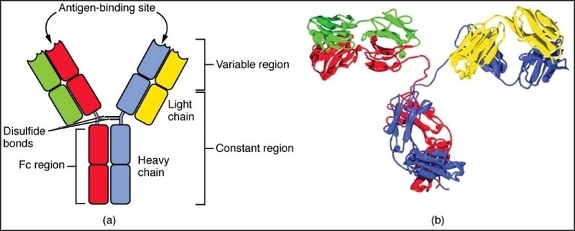
Heart Structure Diagram Image

Heart Structure Diagram Image Diagram - Heart Structure Diagram Image Chart - Human anatomy diagrams and charts explained. This anatomy system diagram depicts Heart Structure Diagram Image with parts and labels. Best diagram to help learn about health, human body and medicine.

Heart Structure Diagram Image

Structure Of The Heart Image

The heart is the organ that helps supply blood and oxygen to all parts of the body. It is divided by a partition (or septum) into two halves, and the halves are in turn divided into four chambers. The heart is situated within the chest cavity and surrounded by a fluid-filled sac called the pericardium.
WebMD’s Heart Anatomy Page provides a detailed image of the heart and provides information on heart conditions, tests, and treatments. Skip to main content Check Your Symptoms Find A Doctor Find A Dentist Connect to Care Find Lowest Drug Prices

Structure Of The Heart Image Diagram - Structure Of The Heart Image Chart - Human anatomy diagrams and charts explained. This anatomy system diagram depicts Structure Of The Heart Image with parts and labels. Best diagram to help learn about health, human body and medicine.

Structure Of The Heart Image

Hip Bone Structure Image

1,047 hip bone stock photos and images available, or search for hip bone icon or hip bone 3d to find more great stock photos and pictures.
Bony Structures of the Hip The hip is formed where the thigh bone (femur) meets the three bones that make up the pelvis: the ilium, the pubis (pubic bone) and the ischium. These three bones converge to form the acetabulum, a deep socket on the outer edge of the pelvis.

Hip Bone Structure Image Diagram - Hip Bone Structure Image Chart - Human anatomy diagrams and charts explained. This anatomy system diagram depicts Hip Bone Structure Image with parts and labels. Best diagram to help learn about health, human body and medicine.

Hip Bone Structure Image

Structure Of The Cardiovascular System Image

Cardiovascular System: Structure & Function. The cardiovascular system or circulatory system is a system which moves nutrients, gases and wastes between cells, helps fight diseases, and transports blood throughout the body (Circulatory System). The main components of the human cardiovascular system include the heart, blood,…
Cardiovascular system – Histology 10 Cardiovascular system Blood circulates throughout the body in blood vessels, propelled by the pumping action of the heart. Blood vessels form a continuous path for blood flow that starts and ends at the heart.

Structure Of The Cardiovascular System Image Diagram - Structure Of The Cardiovascular System Image Chart - Human anatomy diagrams and charts explained. This anatomy system diagram depicts Structure Of The Cardiovascular System Image with parts and labels. Best diagram to help learn about health, human body and medicine.

Structure Of The Cardiovascular System Image

Structure Of The Liver Image

A layer of fibrous tissue called Glisson’s capsule covers the outside of the liver. This capsule is further covered by the peritoneum, a membrane that forms the lining of the abdominal cavity. This helps hold the liver in place and protects it from physical damage. Unlike most organs, the liver has two major sources of blood.
4,742 human liver anatomy stock photos and images available, or start a new search to explore more stock photos and images.

Structure Of The Liver Image Diagram - Structure Of The Liver Image Chart - Human anatomy diagrams and charts explained. This anatomy system diagram depicts Structure Of The Liver Image with parts and labels. Best diagram to help learn about health, human body and medicine.

Structure Of The Liver Image

Diagram Of Bone Structure Image

Microscopic Structure of Bones. The skeleton makes up about 30-40% of an adult’s body mass. The skeleton’s mass is made up of nonliving bone matrix and many tiny bone cells. Roughly half of the bone matrix’s mass is water, while the other half is collagen protein and solid crystals of calcium carbonate and calcium phosphate.
5,814 male human anatomy diagram stock photos and images available, or start a new search to explore more stock photos and images. Male silhouette (contour) on white background, vector. Male, female and children’s silhouette on white background,…

Diagram Of Bone Structure Image Diagram - Diagram Of Bone Structure Image Chart - Human anatomy diagrams and charts explained. This anatomy system diagram depicts Diagram Of Bone Structure Image with parts and labels. Best diagram to help learn about health, human body and medicine.

Diagram Of Bone Structure Image

Human Bone Structure Image

67,060 human bone stock photos and images available, or search for human bone marrow or human bone structure to find more great stock photos and pictures.
Abstract image human body in the form of a starry sky or space, consisting of points, lines, and shapes in the form of planets, stars and the universe. Low poly vector background. human body anatomy stock illustrations Abstract image human body in the form of a starry sky or space,…

Human Bone Structure Image Diagram - Human Bone Structure Image Chart - Human anatomy diagrams and charts explained. This anatomy system diagram depicts Human Bone Structure Image with parts and labels. Best diagram to help learn about health, human body and medicine.

Human Bone Structure Image

Spine Structure Diagram Image

Spine Diagram A diagram of a human female spine showing a side view of the vertebra of the spinal cord within the the body. This is an editable EPS 10 vector illustration with CMYK color space. spinal cord diagram stock illustrations A diagram of a human female spine showing a side view of the vertebra of the spinal cord within the the body.
Browse 2,848 spinal cord diagram stock photos and images available, or search for nervous system or spine illustration to find more great stock photos and pictures. Numbering order of the vertebrae of the human spinal column. Vertebral column: cervical, thoracic and lumbar spine, sacrum and coccyx.
A retro style diagram of the human spine showing the side view with different regions and vertebrae labelled. This is an editable EPS 10 vector illustration with CMYK color space. Computer generated image of spinal column inside gray outline of man, with anatomical labeling, side view on white background.

Spine Structure Diagram Image Diagram - Spine Structure Diagram Image Chart - Human anatomy diagrams and charts explained. This anatomy system diagram depicts Spine Structure Diagram Image with parts and labels. Best diagram to help learn about health, human body and medicine.

Spine Structure Diagram Image

Body Bone Structure Image

Browse 67,060 human bone stock photos and images available, or search for human bone marrow or human bone structure to find more great stock photos and pictures.
Bone Structure. 1 Gross Anatomy of Bone. The structure of a long bone allows for the best visualization of all of the parts of a bone ( (Figure) ). A long bone has two … 2 Bone Markings. 3 Bone Cells and Tissue. 4 Compact and Spongy Bone.
Microscopic Structure of Bones. The skeleton makes up about 30-40% of an adult’s body mass. The skeleton’s mass is made up of nonliving bone matrix and many tiny bone cells. Roughly half of the bone matrix’s mass is water, while the other half is collagen protein and solid crystals of calcium carbonate and calcium phosphate.

Body Bone Structure Image Diagram - Body Bone Structure Image Chart - Human anatomy diagrams and charts explained. This anatomy system diagram depicts Body Bone Structure Image with parts and labels. Best diagram to help learn about health, human body and medicine.

Body Bone Structure Image

Muscle Structure Diagram

Anatomy of the Muscular System Chapter 10281 Figure 10-3Structure of a muscle organ. Note that the connective tissue coverings, the epimysium, perimy- sium, and endomysium, are continuous with each other and with the tendon. Note also that muscle fibers are held together by the perimysium in groups called fascicles.
Tibialis Anterior Muscle Tibialis Posterior Muscle Vastus Lateralis Muscle Vastus Medialis Muscle GENERAL Cross-Sections of Muscles of the Arm and Leg Muscle Cell Types Pronation/Supination Push/Pull Muscles Change Current View Angle Muscular System (Male View) Muscular System (Male Posterior View) Muscular System (Posterior View)
And, together with the scaffolding provided by the skeleton, muscles also determine the form and contours of our body. Contraction of individual muscle cells is ultimately re- sponsible for purposeful movement. In Chapter 11 the phys- iology of muscular contraction is discussed.

Muscle Structure Diagram Diagram - Muscle Structure Diagram Chart - Human anatomy diagrams and charts explained. This anatomy system diagram depicts Muscle Structure Diagram with parts and labels. Best diagram to help learn about health, human body and medicine.

muscle structure diagram

Lymph Node Structure Diagram

Lymph node is small, oval or bean-shaped body. It is a collection of lymphoid tissue enclosed in a connective tis­sue capsule and lying along the lymphatic stream. From the capsule, strands of tissue pass into the substance of the node and are known as the connective tissue trabeculae. In the human lymphatic nodes the trabeculae are ill-de­fined.
Lymph nodes are important structures of the lymphatic system of the thorax and abdomen. They are small, oval or kidney-shaped encapsulated centres of antigen presentation and lymphocyte activation, differentiation and proliferation. They vary in size from 0.1 to 2.5 cm in length.
The lymphoide tissues in the nodes break down materials which have been filtered off such as microorganisms, tumor cells and cells damaged by inflammation. Lymphocyte develops from the reticular and lymphoid tissue in the nodes. Antibodies and antitoxins are also formed by the cells of lymph nodes.

Lymph Node Structure Diagram Diagram - Lymph Node Structure Diagram Chart - Human anatomy diagrams and charts explained. This anatomy system diagram depicts Lymph Node Structure Diagram with parts and labels. Best diagram to help learn about health, human body and medicine.

lymph node structure diagram