Heart Right Atrium Image

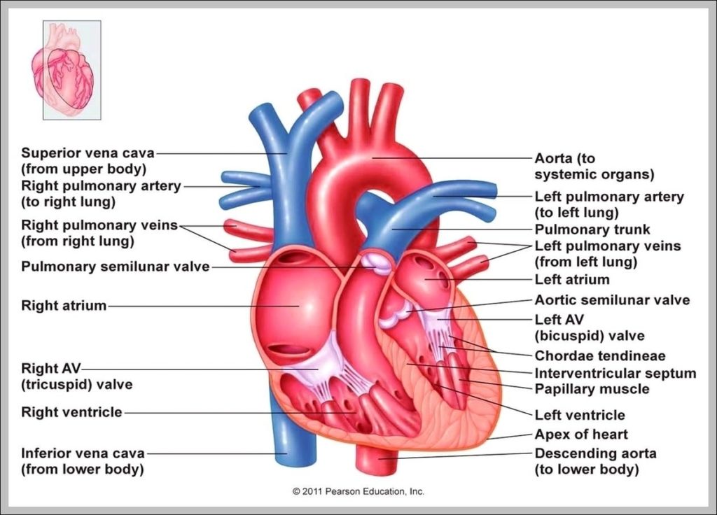
The right atrium is one of the four chambers of the heart. The heart is comprised of two atria and two ventricles. Blood enters the heart through the two atria and exits through the two ventricles. Deoxygenated blood enters the right atrium through the inferior and superior vena cava.
On frontal CXR the findings are consistent with a large right atrium characterized by the globular shape of the heart, large right heart border occupying more than 50% of the cardiovascular height. DOMINANT RIGHT ATRIAL ENLARGEMENT.

Heart Right Atrium Image Diagram - Heart Right Atrium Image Chart - Human anatomy diagrams and charts explained. This anatomy system diagram depicts Heart Right Atrium Image with parts and labels. Best diagram to help learn about health, human body and medicine.

Heart Right Atrium Image

Right Lymphatic Duct Image

Right Lymphatic Duct. The right lymphatic duct, also called the right thoracic duct, is about 1.25 cm long. It drains lymphatic fluid from the right thoracic cavity (this is the section of the trunk on the upper right side), the right arm, and from the right side of the neck and the head.
The human body contains two main types of lymph ducts: the thoracic duct and the right lymph duct. The first primary lymph duct runs along the abdomen to the neck.

Right Lymphatic Duct Image Diagram - Right Lymphatic Duct Image Chart - Human anatomy diagrams and charts explained. This anatomy system diagram depicts Right Lymphatic Duct Image with parts and labels. Best diagram to help learn about health, human body and medicine.

Right Lymphatic Duct Image