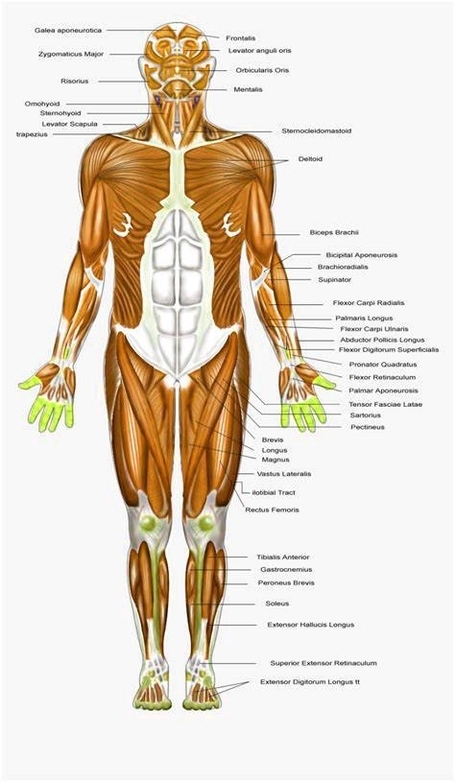
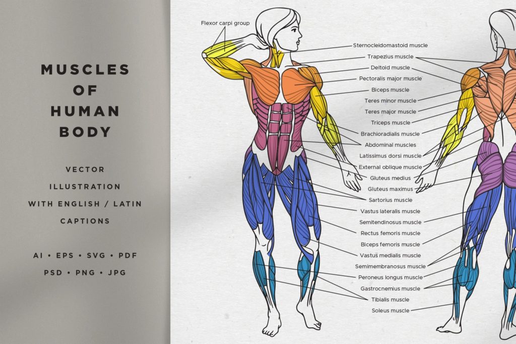
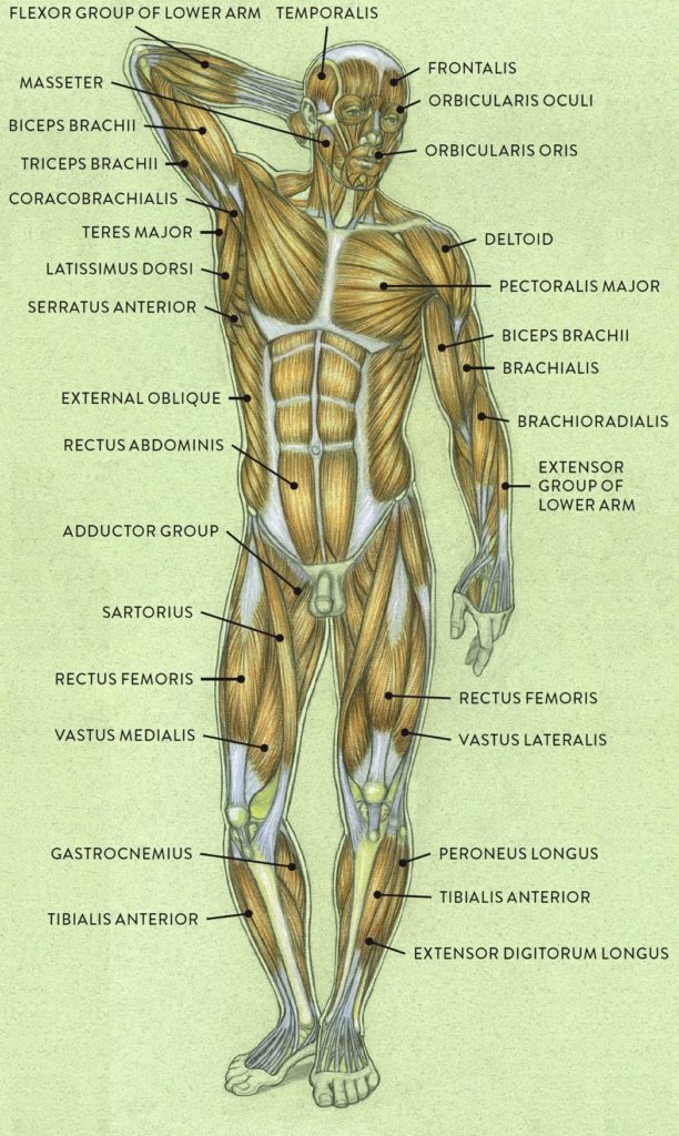
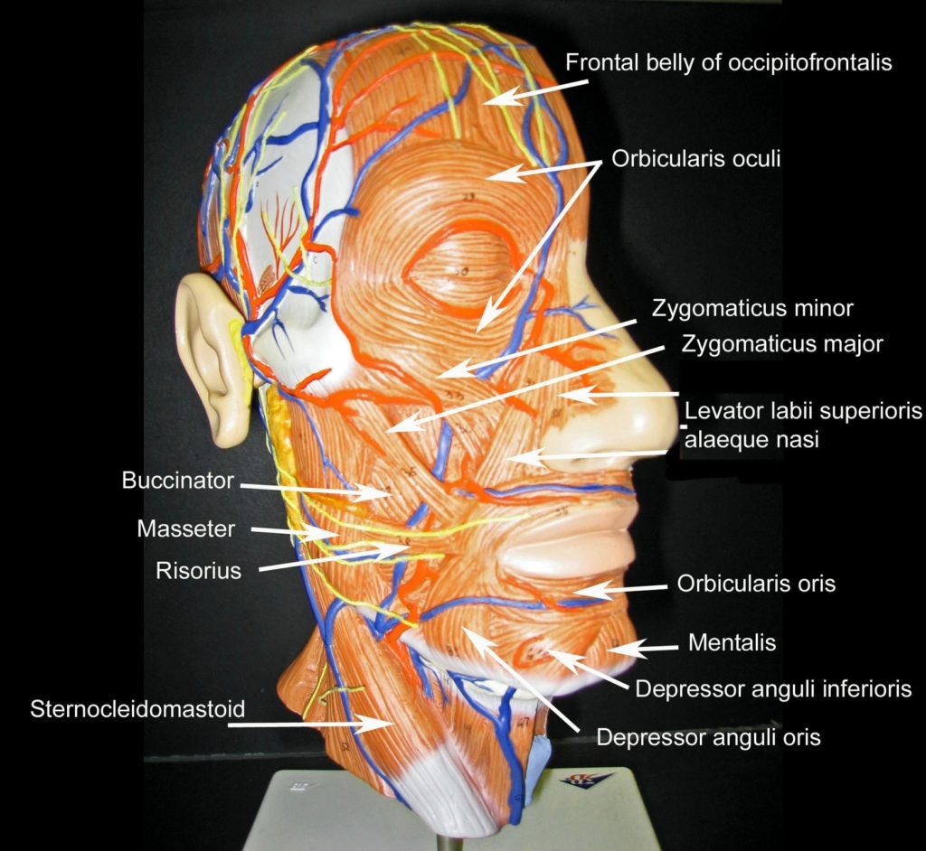
Major Muscles of the Human Body The human body is a complex system that comprises more than 600 muscles. These muscles are broadly divided into three types: 1. Skeletal Muscle:…
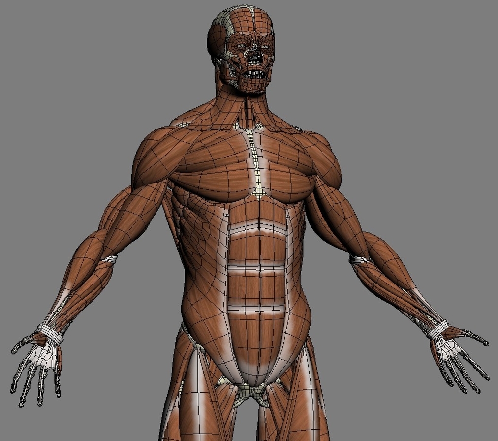
The upper body muscles are a complex network that enables a wide range of movements and provides the strength for many functional tasks. They can be broadly categorized into muscles…
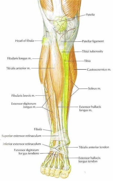
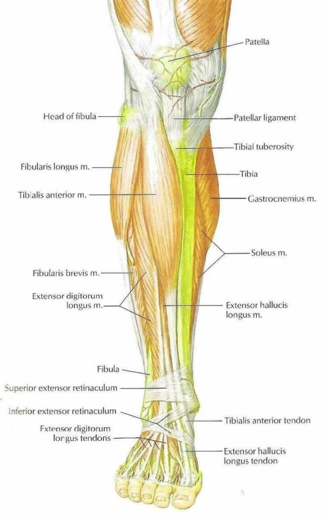
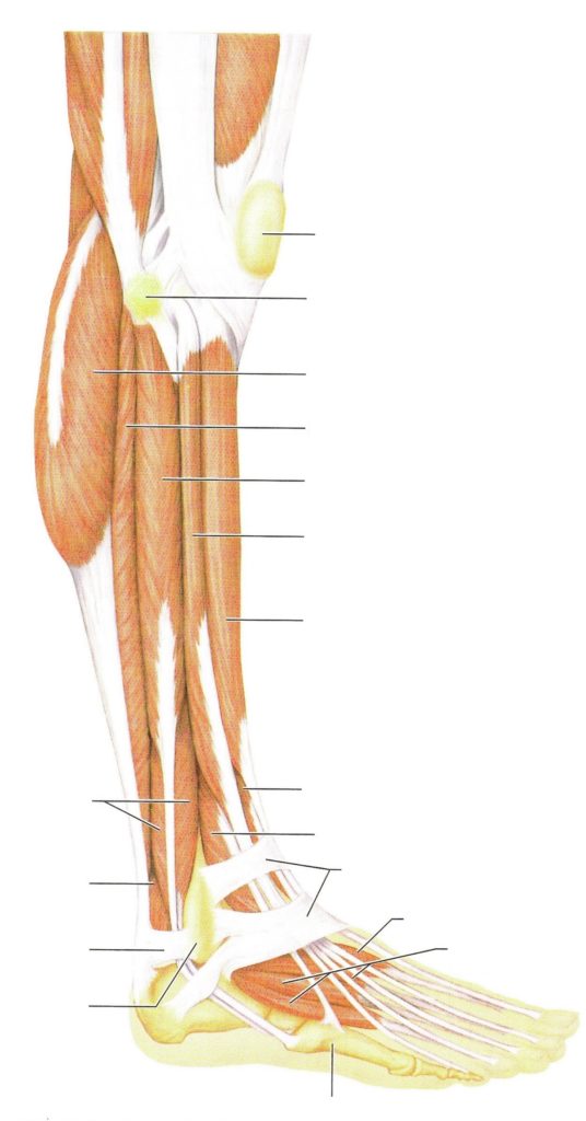
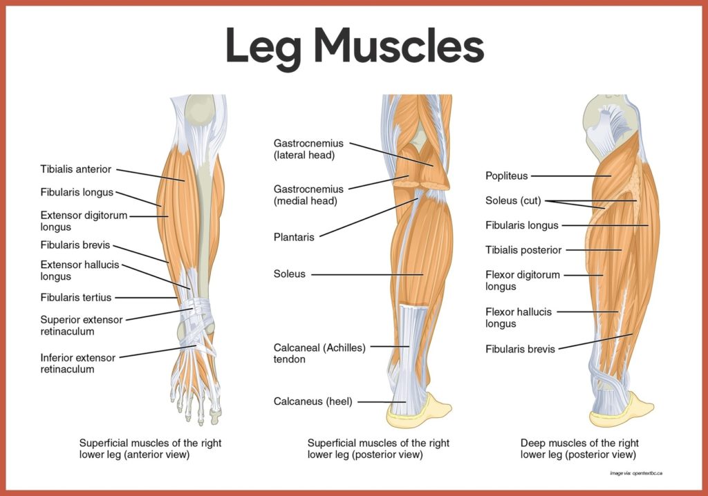
Lower Leg Muscles Study The lower leg, anatomically defined as the region of the lower limb below the knee, is a complex structure that plays a crucial role in movements…
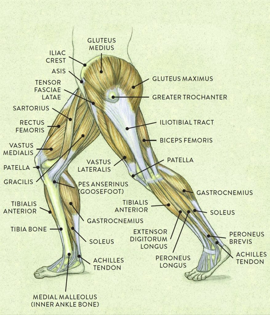
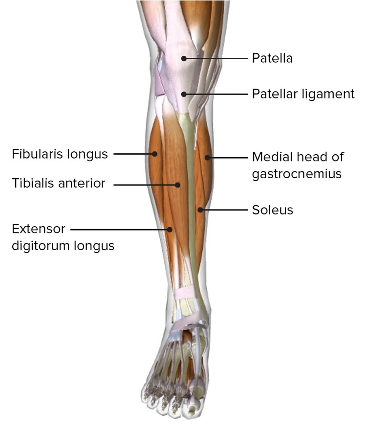
Anatomy of Leg Muscles The leg, anatomically defined as the region of the lower limb from the knee to the ankle, is composed of various muscles that enable movements like…
The lower leg, anatomically defined as the region of the lower limb below the knee, is a complex structure that plays a crucial role in weight-bearing activities such as walking,…
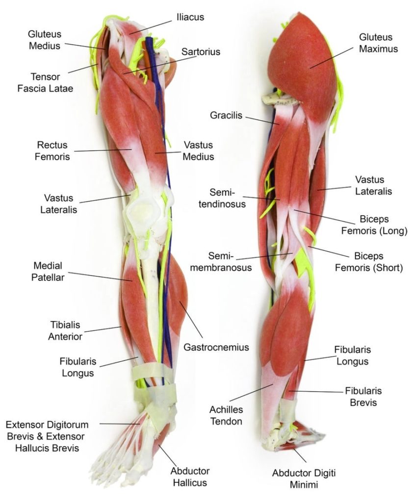
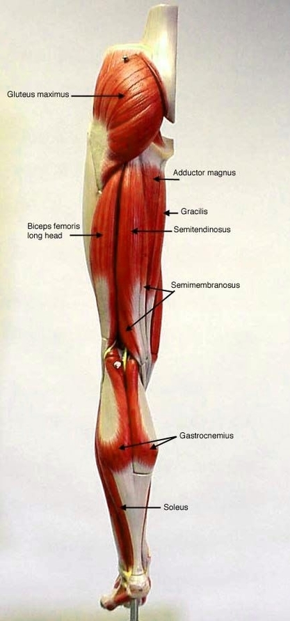
The human leg, anatomically defined as the region of the lower limb from the knee to the ankle, is a complex structure composed of numerous muscles that work in harmony…
Human Body Muscles Examined The human body is a complex system that comprises more than 600 muscles. These muscles, which are pieces of soft tissue, are involved in everything from…
the anterior compartment muscles of the leg. These muscles play a crucial role in movement, stability, and overall function of the lower limb. Without further ado, let's explore their anatomy,…
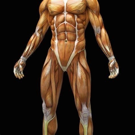
Understanding Leg Muscles and Their Importance in Gym Workouts The leg muscles are a complex group of muscles that not only support the body's weight but also play a crucial…
The human torso, also known as the trunk, is the central part of the body to which the head and the limbs are attached. It houses all the vital organs…
abdominal muscles. These essential muscles play a crucial role in maintaining stability, supporting movement, and protecting vital organs. Without further ado, let's explore the intricacies of these remarkable structures. ##…
The lower back muscles, also known as the lumbar region muscles, play a crucial role in maintaining the structural integrity of the body. They provide support to the spine, aid…
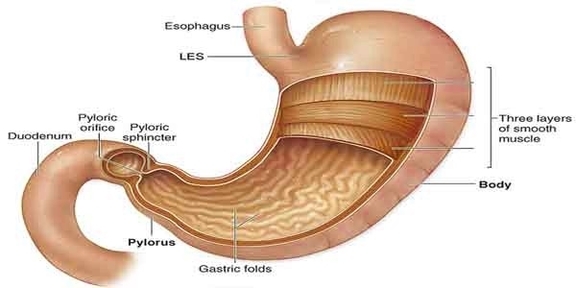
stomach muscles. The stomach, that resilient organ responsible for digesting our meals, is more than just a sack where food temporarily resides. It's a complex structure with layers of muscles…
Label Muscles Worksheet A Label Muscles Worksheet is an educational tool designed to help students learn about the muscular system of the human body. It typically includes diagrams of the…
Human Body Muscles The human body is a complex system that relies on muscles for movement, posture, and balance. There are three major types of muscles in the human body:…
Stomach Muscles and Digestive System Anatomy The stomach is a muscular organ that plays a crucial role in the digestive system. It is part of the gastrointestinal (GI) tract, which…
Human Body Muscles: An Overview The human body is a complex system, and one of its most essential components is the muscular system. Comprising more than 600 muscles, this system…
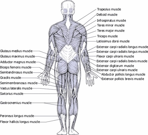
Human Muscles: Back View The human back, a complex structure comprising bones, muscles, and nerves, plays a crucial role in body support, movement, and protection of vital organs. pine The…
The human leg, a marvel of biological engineering, is powered by a complex network of muscles. These muscles, working in harmony, enable us to perform a wide range of movements,…
Leg Muscles Anatomy Study The leg muscles, anatomically defined as the region of the lower limb below the knee, are organized into three compartments: anterior, posterior, and lateral. Anterior (Dorsiflexor)…