A Skeleton With Labelshuman Skeleton Diagram With Labels Explanation

The human skeleton, an intricate internal framework, provides essential support, protection, and mobility for our bodies. Comprising numerous individual bones and cartilages, it forms the architectural basis upon which our muscles, organs, and other soft tissues rely. the human skeleton, exploring its structure, functions, and significance.

## Anatomy of the Human Skeleton

1. Axial Skeleton:
– The vertebral column, commonly known as the spine, constitutes the central axis of the axial skeleton. It serves as the primary support for the trunk and head.
– Much of the skull also falls within the axial skeleton. The skull houses the brain, sensory organs (such as the eyes and ears), and the intricate network of cranial nerves.
– Additionally, the visceral subdivision—which includes the lower jaw, parts of the upper jaw, and the branchial arches (including the hyoid bone)—is part of the axial skeleton.

2. Appendicular Skeleton:
– The appendicular skeleton encompasses the bones and cartilages of the limbs, as well as the pectoral (shoulder) girdle and the pelvic (hip) girdle.
– It facilitates movement, allowing us to walk, run, grasp objects, and perform various activities.

## Functions of the Skeleton

1. Support:
– The skeleton provides a sturdy framework that maintains the body’s shape and prevents it from collapsing under its own weight.
– The vertebral column, akin to the notochord in lower organisms, plays a crucial role in supporting the trunk.

2. Protection:
– Bones shield vital organs from external forces. For instance:
– The skull protects the brain.
– The rib cage safeguards the heart and lungs.
– The vertebral column shields the spinal cord.

3. Motion:
– The appendicular skeleton enables movement by serving as attachment points for muscles.
– Joints—where bones meet—allow for flexibility and coordinated motion.
– Muscles pull on bones, creating the mechanical force necessary for locomotion.

## Notable Bones and Their Functions

1. Femur:
– The thigh bone is the longest and strongest bone in the human body.
– It supports body weight during standing, walking, and running.

2. Scapula (Shoulder Blade):
– The scapula anchors muscles that move the arm and shoulder.
– It provides stability for arm movements.

3. Humerus:
– The upper arm bone connects the shoulder to the elbow.
– It allows for arm flexion, extension, and rotation.

4. Radius and Ulna:
– These forearm bones enable wrist rotation and hand movements.
– The radius is on the thumb side, while the ulna is on the pinky side.

5. Tibia and Fibula:
– The shinbones form the lower leg.
– The tibia bears most of the body weight during standing.

6. Pelvic Bones:
– The pelvis consists of the ilium, ischium, and pubis.
– It supports the abdominal organs and provides attachment for leg muscles.

## Labeling the Skeleton

– To understand the human skeleton better, consider using labeled diagrams and interactive resources.
– These resources help identify individual bones, their locations, and their relationships to other structures.
– Whether in the classroom or as a fun activity at home, exploring the science of the human body through skeletal labeling enhances our appreciation for this remarkable framework.

In summary, the human skeleton—composed of bones, cartilages, ligaments, and tendons—serves as an intricate scaffold. It not only supports our bodies but also ensures our survival, allowing us to move, protect our vital organs, and thrive in our dynamic world..

A Skeleton With Labelshuman Skeleton Diagram With Labels Explanation Diagram - A Skeleton With Labelshuman Skeleton Diagram With Labels Explanation Chart - Human anatomy diagrams and charts explained. This anatomy system diagram depicts A Skeleton With Labelshuman Skeleton Diagram With Labels Explanation with parts and labels. Best diagram to help learn about health, human body and medicine.

A Skeleton With Labelshuman Skeleton Diagram With Labels Explanation

A Skeleton With Labelshuman Skeleton Diagram With Labels With Labels

The human skeleton, an intricate internal framework, serves as the architectural support for our bodies. Comprising numerous individual bones and cartilages, it provides the essential structure upon which our muscles, organs, and other soft tissues rely. the human skeleton.

## Axial Skeleton: The Core Support

1. Vertebral Column (Spine):
– The vertebral column, akin to the notochord in lower organisms, forms the central axis of the axial skeleton. It consists of a series of 33 vertebrae, each with distinct features.
– These vertebrae are categorized into five regions: cervical (neck), thoracic (mid-back), lumbar (lower back), sacral (fused into the sacrum), and coccygeal (tailbone).
– The spine provides support, flexibility, and protection for the spinal cord.

2. Skull:
– The skull houses the brain and sensory organs.
– It comprises 22 bones, including the cranium (protecting the brain) and the facial bones (forming the visage).
– The mandible (lower jaw) and the hyoid bone (associated with swallowing) are also part of the skull.

3. Visceral Skeleton:
– This subdivision includes the lower jaw, certain elements of the upper jaw, and the branchial arches.
– The hyoid bone, suspended in the neck, plays a role in swallowing and speech.

## Appendicular Skeleton: Mobility and Functionality

1. Pectoral Girdle (Shoulder):
– Comprising the clavicle (collarbone) and scapula (shoulder blade), the pectoral girdle connects the upper limbs to the axial skeleton.
– It allows for arm movement and provides stability.

2. Upper Limbs:
– The upper limbs consist of the humerus (upper arm bone), radius and ulna (forearm bones), and various hand bones.
– These bones facilitate intricate movements, from writing to playing musical instruments.

3. Pelvic Girdle (Hip):
– The pelvis consists of the ilium, ischium, and pubis bones.
– It supports the trunk, transfers weight to the lower limbs, and protects reproductive and digestive organs.

4. Lower Limbs:
– The lower limbs include the femur (thigh bone), tibia and fibula (leg bones), and the intricate bones of the foot.
– These bones enable walking, running, and maintaining balance.

## Functions of the Skeleton

1. Support:
– The skeleton provides a sturdy framework for the body, maintaining its shape and preventing collapse.
– The vertebral column, akin to a resilient pillar, bears the weight of the upper body.

2. Protection:
– Bones shield vital organs. For instance, the skull envelops the brain, and the rib cage safeguards the heart and lungs.

3. Motion:
– Joints between bones allow movement. Some joints, like the ball-and-socket joint in the hip, permit a wide range of motion.
– Muscles attach to bones via tendons, enabling coordinated movement.

In summary, the human skeleton is a remarkable blend of strength, flexibility, and intricate design. It silently supports our every action, from the simplest gesture to the most complex dance. ????

: [Britannica](https://www.britannica.com/science/human-skeleton)
: [Britannica](https://www.britannica.com/facts/human-skeleton)
: [Britannica](https://www.britannica.com/summary/skeleton)
: [Wikipedia](https://en.wikipedia.org/wiki/Human_skeleton).
o

A Skeleton With Labelshuman Skeleton Diagram With Labels With Labels Diagram - A Skeleton With Labelshuman Skeleton Diagram With Labels With Labels Chart - Human anatomy diagrams and charts explained. This anatomy system diagram depicts A Skeleton With Labelshuman Skeleton Diagram With Labels With Labels with parts and labels. Best diagram to help learn about health, human body and medicine.

A Skeleton With Labelshuman Skeleton Diagram With Labels With Labels

A Skeleton With Labelshuman Skeleton Diagram With Labels Sample

The human skeleton, an intricate internal framework, serves as the structural backbone for our bodies. Comprising numerous individual bones and cartilages, it provides essential support, protection, and facilitates movement. Let’s delve into the fascinating details of this remarkable system.

## Axial Skeleton
The axial skeleton forms the central axis of the body. It consists of the following components:

1. Vertebral Column (Spine):
– The vertebral column, akin to the notochord in lower organisms, is the primary support for the trunk.
– Comprising 33 vertebrae, it extends from the skull to the coccyx.
– These vertebrae are categorized into five regions: cervical (neck), thoracic (chest), lumbar (lower back), sacral (pelvic), and coccygeal (tailbone).
– Intervertebral discs cushion and allow flexibility between adjacent vertebrae.

2. Skull:
– The skull safeguards the brain and houses sensory organs.
– It consists of 22 bones, including the cranium (protecting the brain) and facial bones (forming the face).
– The mandible (lower jaw) and maxilla (upper jaw) play crucial roles in chewing and speech.

3. Hyoid Bone:
– The hyoid bone, part of the visceral subdivision, lies in the throat.
– It anchors muscles involved in swallowing and speech.

## Appendicular Skeleton
The appendicular skeleton comprises bones and cartilages related to the limbs and their attachment points:

1. Pectoral (Shoulder) Girdle:
– The pectoral girdle includes the clavicle (collarbone) and scapula (shoulder blade).
– These bones connect the upper limbs to the axial skeleton.

2. Pelvic (Hip) Girdle:
– The pelvic girdle consists of the ilium, ischium, and pubis bones.
– It supports the lower limbs and protects pelvic organs.

3. Upper Limbs:
– Each upper limb comprises the humerus (upper arm bone), radius, and ulna (forearm bones).
– The carpals (wrist bones), metacarpals (palm bones), and phalanges (finger bones) complete the upper limb.

4. Lower Limbs:
– The lower limb includes the femur (thigh bone), patella (kneecap), tibia, and fibula (leg bones).
– The tarsals (ankle bones), metatarsals (foot bones), and phalanges (toe bones) form the rest of the limb.

## Functions of the Skeleton
1. Support:
– The skeleton provides a sturdy framework, maintaining body shape and preventing collapse.
– The vertebral column, especially, ensures upright posture.

2. Protection:
– Bones shield vital organs. For instance, the skull protects the brain, and the ribcage guards the heart and lungs.

3. Motion:
– Joints, where bones meet, allow movement.
– Muscles attach to bones via tendons, enabling locomotion.

In summary, the human skeleton, with its intricate divisions, plays a multifaceted role—providing support, safeguarding organs, and allowing us to move through life’s journey. ???????

: [Britannica](https://www.britannica.com/science/human-skeleton).
o

A Skeleton With Labelshuman Skeleton Diagram With Labels Sample Diagram - A Skeleton With Labelshuman Skeleton Diagram With Labels Sample Chart - Human anatomy diagrams and charts explained. This anatomy system diagram depicts A Skeleton With Labelshuman Skeleton Diagram With Labels Sample with parts and labels. Best diagram to help learn about health, human body and medicine.

A Skeleton With Labelshuman Skeleton Diagram With Labels Sample

A Skeleton With Labelshuman Skeleton Diagram With Labels Visual

The human skeleton, an intricate framework of bones and cartilage, provides essential support, protection, and mobility for our bodies. Comprising 206 bones in adults, the skeleton is a remarkable system that plays a crucial role in maintaining our physical form and enabling various functions.

## 1. Structure and Composition:
– The human skeleton consists of two main divisions: the axial skeleton and the appendicular skeleton.
– Axial Skeleton:
– Forms the central axis of the body.
– Includes the skull, vertebral column, and rib cage.
– The skull comprises the cranium (protecting the brain) and the facial bones.
– The vertebral column (or spine) consists of 33 vertebrae, providing flexibility and support.
– The rib cage safeguards vital organs like the heart and lungs.
– Appendicular Skeleton:
– Attaches to the axial skeleton.
– Includes the limbs, shoulder girdle, and pelvic girdle.
– The shoulder girdle connects the upper limbs to the axial skeleton.
– The pelvic girdle supports the lower limbs and protects reproductive organs.

## 2. Bone Types:
– Bones are classified into five types based on shape and function:
1. Long Bones:
– Longer than they are wide.
– Examples: femur (thigh bone), humerus (upper arm bone).
2. Short Bones:
– Cube-shaped.
– Provide stability and support.
– Examples: carpals (wrist bones), tarsals (ankle bones).
3. Flat Bones:
– Thin and flat.
– Protect internal organs and provide attachment points.
– Examples: scapula (shoulder blade), sternum (breastbone).
4. Irregular Bones:
– Complex shapes.
– Serve specific functions.
– Examples: vertebrae, facial bones.
5. Sesamoid Bones:
– Develop within tendons.
– Reduce friction and enhance mechanical advantage.
– Example: patella (kneecap).

## 3. Bone Tissue:
– Bones consist of compact bone (dense and strong) and spongy bone (trabecular bone with a porous structure).
– Osteocytes, embedded in the bone matrix, maintain bone health.
– Haversian canals contain blood vessels and nerves, ensuring nutrient supply.

## 4. Joints:
– Joints connect bones and allow movement.
– Types of joints:
– Fibrous Joints: Immovable (e.g., sutures in the skull).
– Cartilaginous Joints: Slightly movable (e.g., intervertebral discs).
– Synovial Joints: Freely movable (e.g., ball-and-socket, hinge joints).
– Examples: shoulder joint, hip joint, knee joint.

## 5. Bone Development and Growth:
– Ossification:
– Process of bone formation.
– Intramembranous ossification: Forms flat bones directly from mesenchymal tissue.
– Endochondral ossification: Converts cartilage into bone (long bones).
– Bone Remodeling:
– Ongoing process of resorption and deposition.
– Maintains bone strength and adapts to stress.

## 6. Common Bones:
– Skull Bones:
– Cranium (frontal, parietal, temporal, occipital, sphenoid, ethmoid).
– Facial bones (maxilla, mandible, zygomatic, nasal, etc.).
– Vertebral Column:
– Cervical (neck), thoracic (mid-back), lumbar (lower back), sacral, and coccygeal vertebrae.
– Upper Limb Bones:
– Humerus, radius, ulna, carpals, metacarpals, and phalanges.
– Lower Limb Bones:
– Femur, tibia, fibula, patella, tarsals, metatarsals, and phalanges.

## 7. Clinical Significance:
– Osteoporosis: Bone density loss.
– Fractures: Breaks in bones.
– Arthritis: Joint inflammation.
– Scoliosis: Abnormal spinal curvature.

In summary, the human skeleton is a marvel of engineering, providing both strength and flexibility. Its intricate design ensures our survival, movement, and protection throughout life..

A Skeleton With Labelshuman Skeleton Diagram With Labels Visual Diagram - A Skeleton With Labelshuman Skeleton Diagram With Labels Visual Chart - Human anatomy diagrams and charts explained. This anatomy system diagram depicts A Skeleton With Labelshuman Skeleton Diagram With Labels Visual with parts and labels. Best diagram to help learn about health, human body and medicine.

A Skeleton With Labelshuman Skeleton Diagram With Labels Visual

A Skeleton With Labelshuman Skeleton Diagram With Labels Graphic

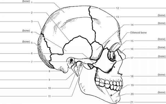
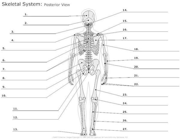
The human skeleton, an intricate internal framework, serves as the architectural basis for our bodies. Comprising numerous individual bones and cartilages, it provides structural support, protection, and facilitates movement. Let’s delve into the fascinating details of this remarkable system.

## Axial Skeleton: The Core Support

1. Vertebral Column (Spine):
– The vertebral column, akin to the notochord in lower organisms, forms the central axis of the axial skeleton. It consists of 33 vertebrae, grouped into five regions: cervical, thoracic, lumbar, sacral, and coccygeal.
– These vertebrae encase and protect the delicate spinal cord, allowing us to stand upright and maintain posture.
– Intervertebral discs cushion the vertebrae, absorbing shocks and enabling flexibility.

2. Skull:
– The skull houses the brain, sensory organs, and the intricate cranial nerves.
– It comprises 22 bones, including the cranium (protecting the brain) and facial bones (forming the eye sockets, nasal cavity, and jaw).
– The hyoid bone, part of the visceral subdivision, supports the tongue and aids in swallowing.

3. Thorax:
– The rib cage safeguards vital organs such as the heart and lungs.
– Twelve pairs of ribs articulate with the thoracic vertebrae, forming the rib cage.
– The sternum (breastbone) connects the ribs anteriorly.

## Appendicular Skeleton: Mobility and Functionality

1. Upper Limbs:
– The pectoral girdle (shoulder) consists of the clavicle (collarbone) and scapula (shoulder blade).
– The humerus, radius, and ulna form the arm and forearm.
– The carpals, metacarpals, and phalanges constitute the hand.

2. Lower Limbs:
– The pelvic girdle (hip) includes the ilium, ischium, and pubis bones.
– The femur, tibia, and fibula create the thigh and leg.
– The tarsals, metatarsals, and phalanges compose the foot.

3. Functions of the Skeleton:
– Support: The skeleton’s primary role is to provide a sturdy framework for the body.
– Protection: Bones shield delicate organs. For instance, the rib cage guards the heart and lungs.
– Motion: Joints between bones allow movement. Some joints, like the ball-and-socket joint in the hip, offer a wide range of motion.
– Hematopoiesis: Certain bones, such as the sternum and pelvis, produce blood cells.

In summary, the human skeleton, with its intricate interplay of bones, cartilage, ligaments, and tendons, is a marvel of design. It upholds our bodies, safeguards vital organs, and enables graceful movement—a silent architect shaping our existence..

A Skeleton With Labelshuman Skeleton Diagram With Labels Graphic Diagram - A Skeleton With Labelshuman Skeleton Diagram With Labels Graphic Chart - Human anatomy diagrams and charts explained. This anatomy system diagram depicts A Skeleton With Labelshuman Skeleton Diagram With Labels Graphic with parts and labels. Best diagram to help learn about health, human body and medicine.

A Skeleton With Labelshuman Skeleton Diagram With Labels Graphic