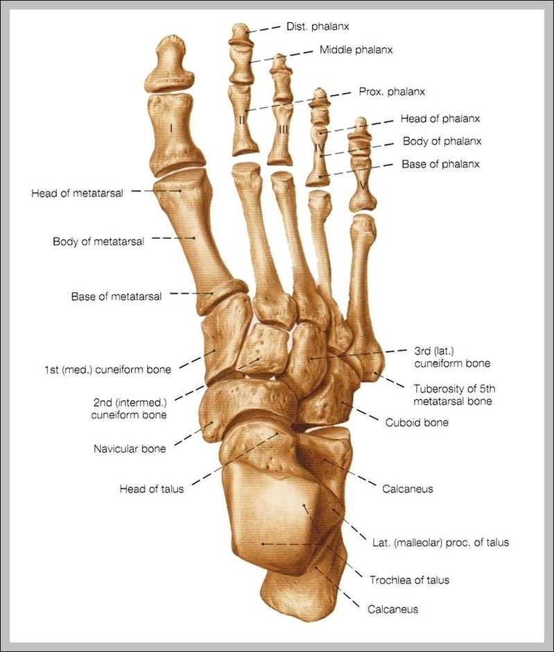
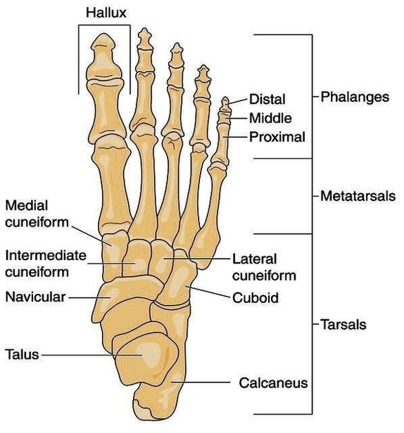
The human foot, a marvel of biological engineering, is one of the most complex structures in the body. It is composed of over 100 moving parts, including 26 bones, 33…
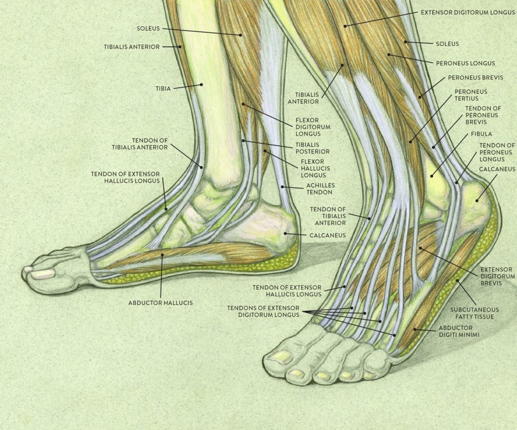
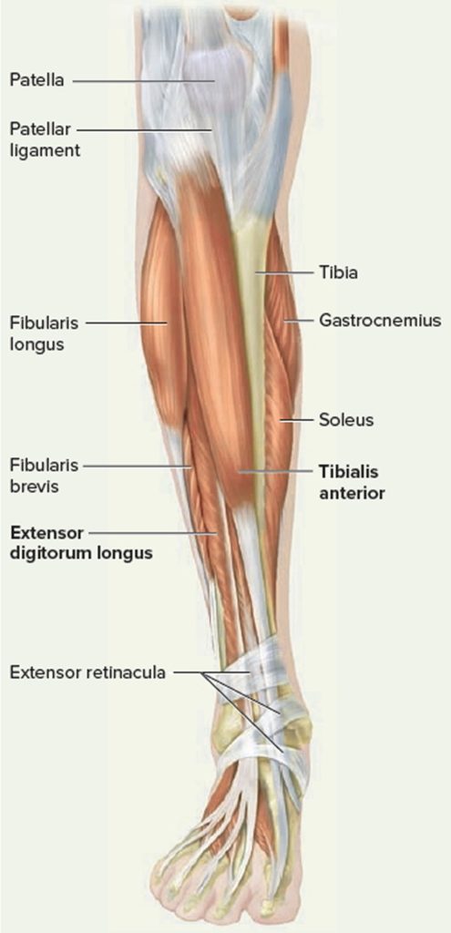
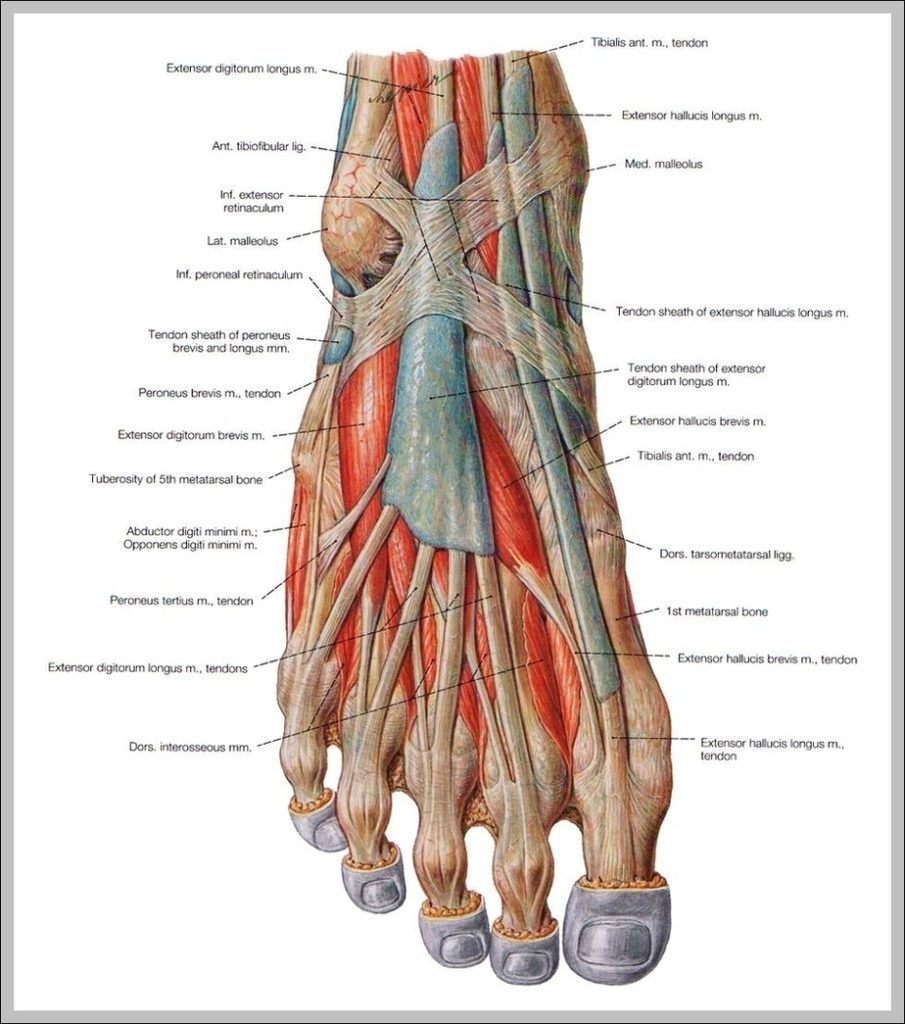
Tendons are thick bands of tissue that connect muscles to bone. When a muscle contracts, the tendon pulls on the bone causing the joint to move. There are a number…
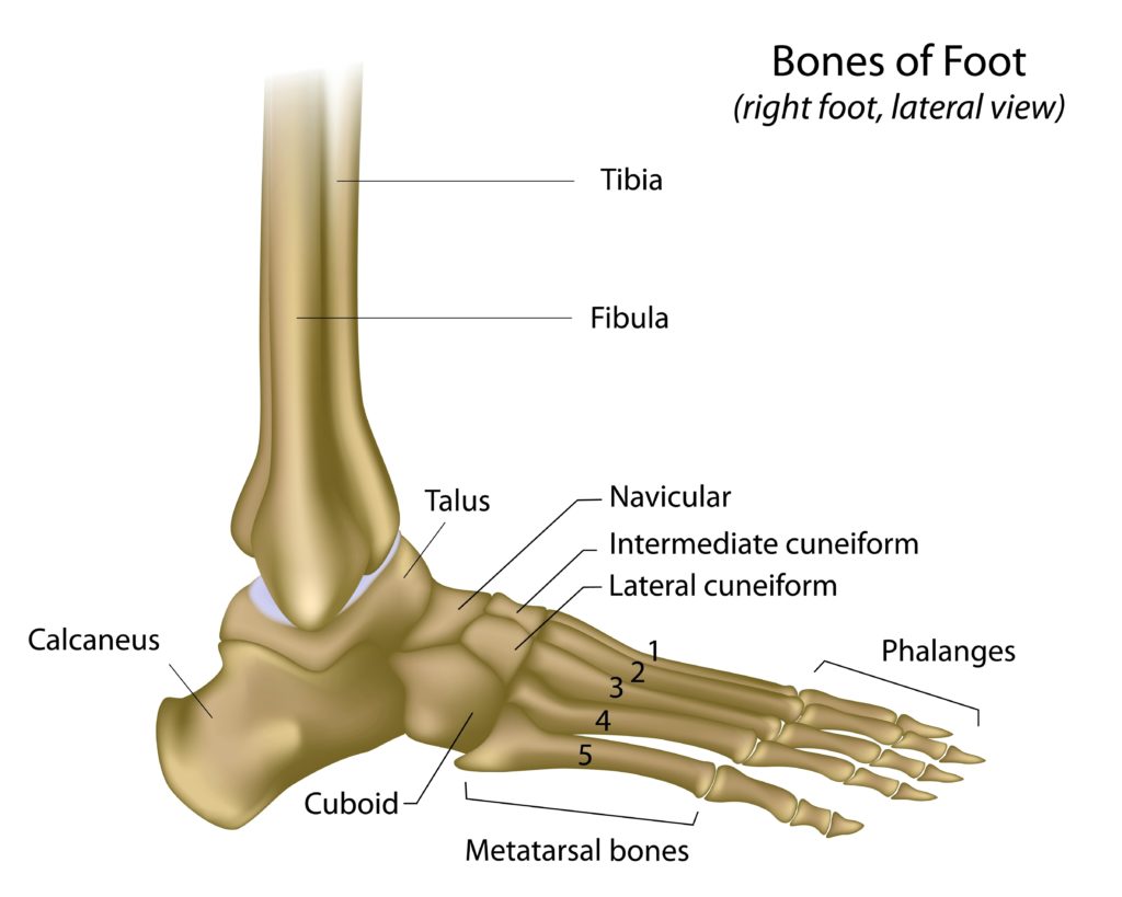
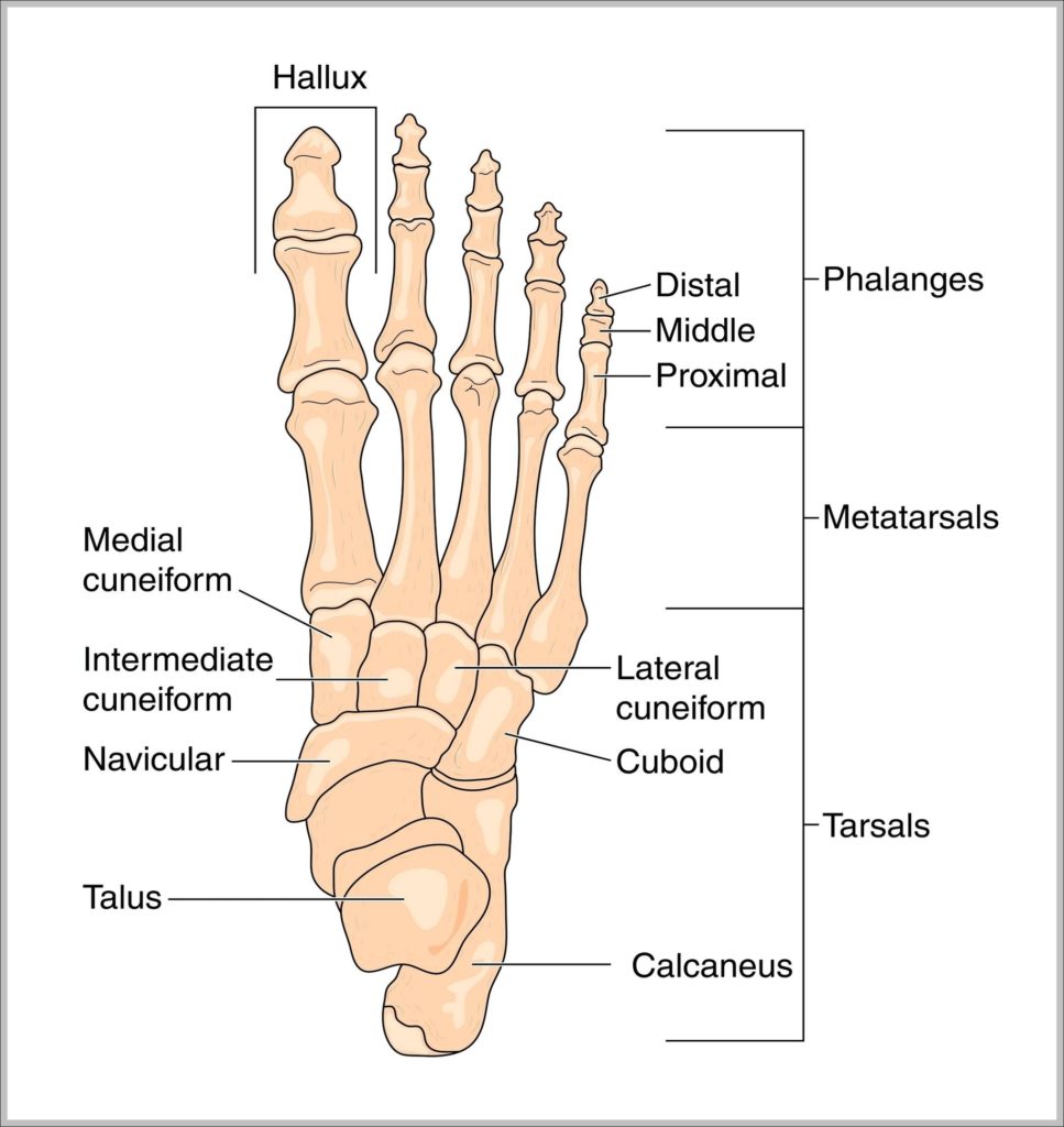
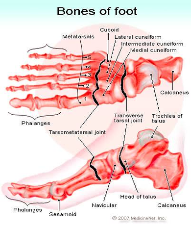
The foot and ankle form a complex system which consists of 28 bones, 33 joints, 112 ligaments, controlled by 13 extrinsic and 21 intrinsic muscles?. The foot is subdivided into…
4,379 human body anatomy female stock photos and images available, or start a new search to explore more stock photos and images. A guide to female anatomy. Female anatomy includes…
80,933 human internal organ stock photos and images available or search for human internal organ icons or human internal organ illustrations to find more great stock photos and pictures. Human…
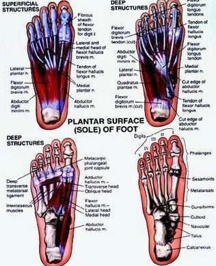
The muscles of the foot can be split into two groups, the extrinsic and intrinsic muscles. The extrinsic foot muscles are found in the lower leg and act to dorsiflex,…
The anatomy of the foot. The foot contains a lot of moving parts - 26 bones, 33 joints and over 100 ligaments. The foot is divided into three sections -…
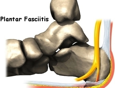
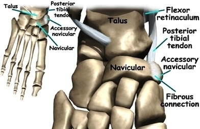
Because there are so many bones in the foot, there are also numerous ligaments connecting them. Some of the main ligaments in the foot are: Plantar fascia ligament: Runs underneath…
Model of a skeletal foot. A highly detailed articulated model of a human foot, with all the bones represented, from the toes to just past the ankle This Part of…
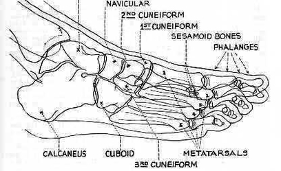
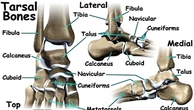
The skeletal structure of the foot is similar to that of the hand but, because the foot bears more weight, it is stronger but less movable. The bones of the…