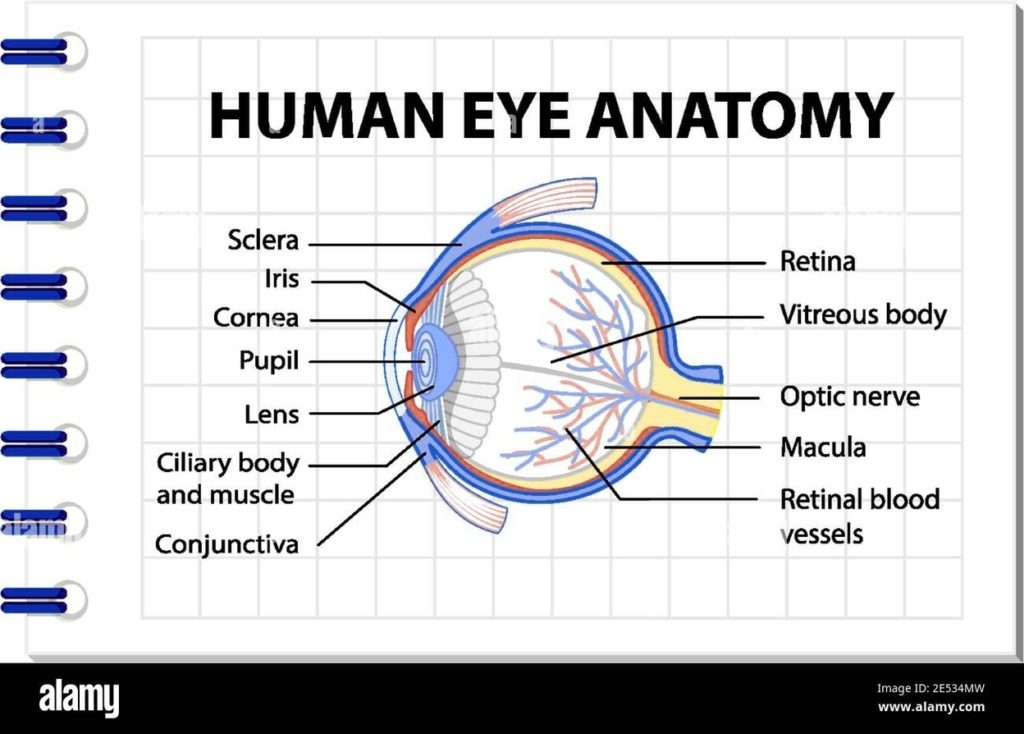
The human eye, a marvel of biological engineering, is a complex organ that plays a crucial role in the perception of visual stimuli. Here's a detailed look at its anatomy:…
The Iris and Eye Structure The human eye is a complex organ that allows us to perceive the world around us. One of its key components is the iris, the…
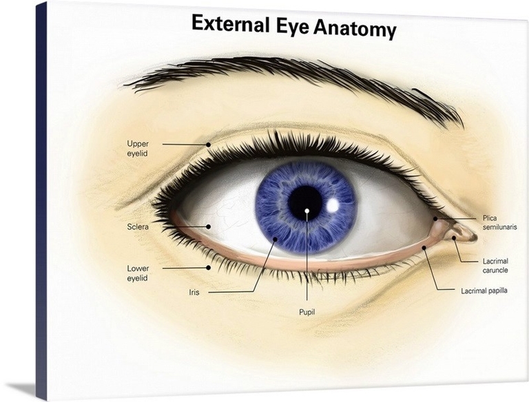
The human eye is a complex organ that allows us to perceive the world around us. The external anatomy of the eye includes several key components, each with a specific…
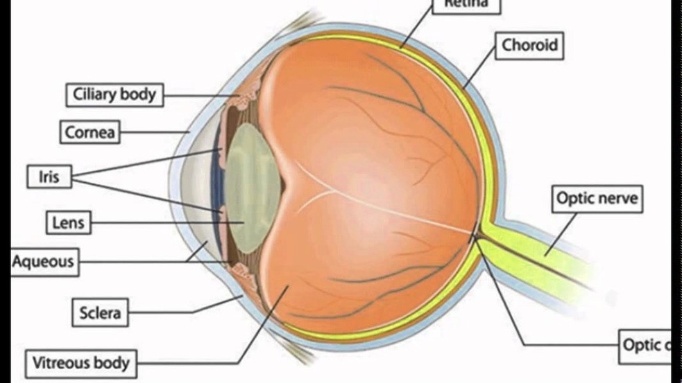
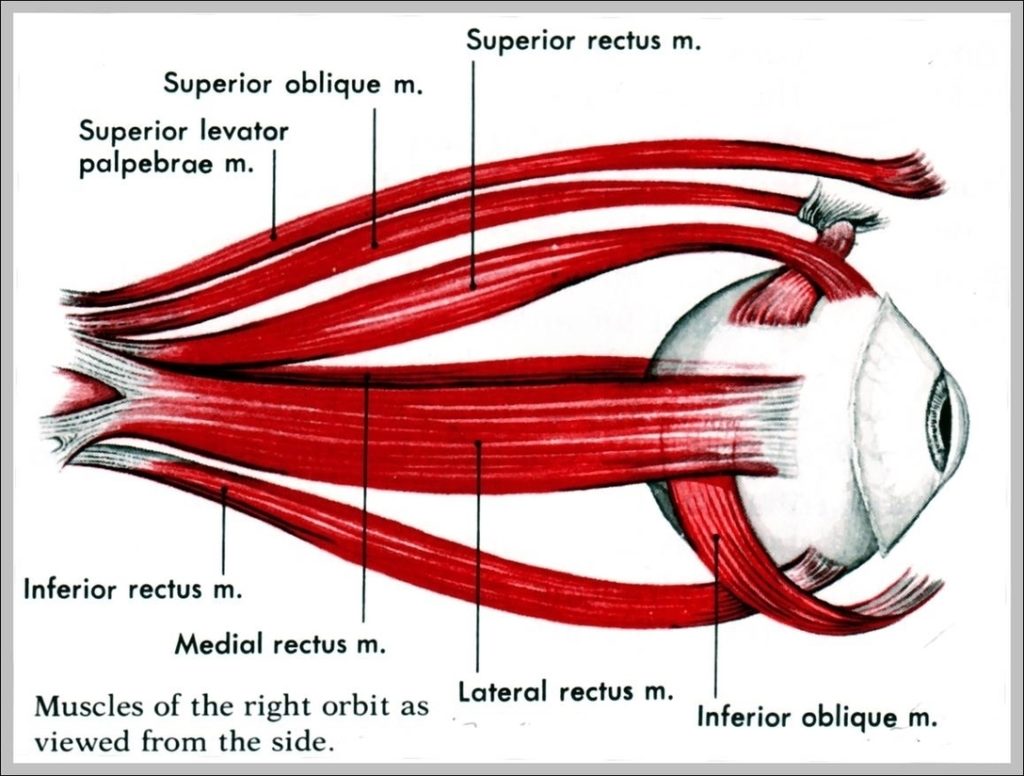
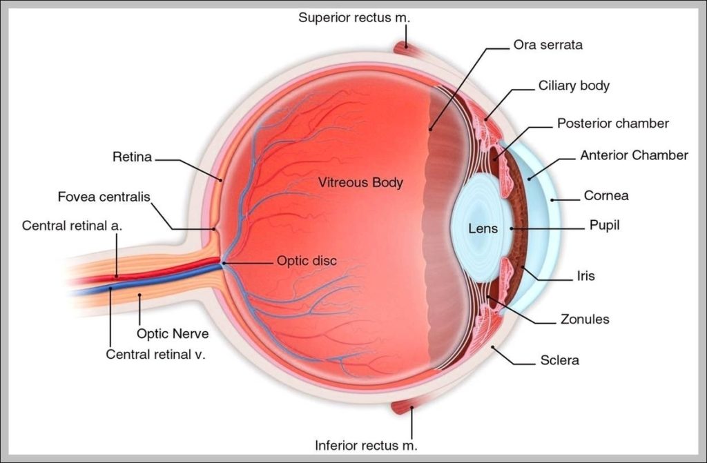
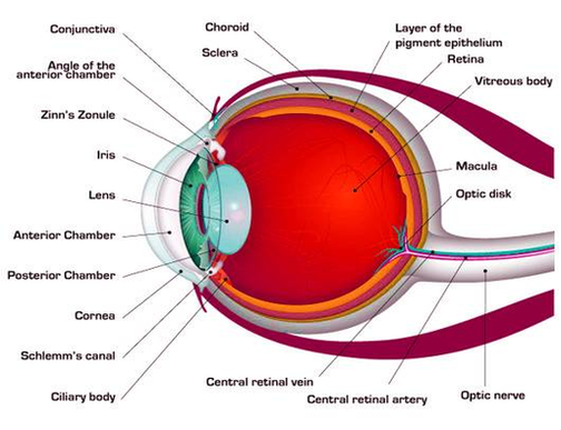
The Human Eye (Eyeball) Diagram, Parts and Pictures. The human eye consists of the eyeball, optic nerve, orbit and appendages (eyelids, extraocular muscles and lacrimal glands). While the eyeball is…
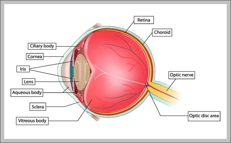
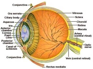
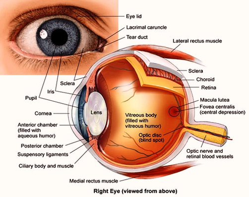
Dimensions:760 x 772 Photo description: This human eye diagram gives an excellent overview of the human eye. The cross section features labeled parts such as the iris, pupil, cornea, lens,…
The Human Eye (Eyeball) Diagram, Parts and Pictures. The human eye consists of the eyeball, optic nerve, orbit and appendages (eyelids, extraocular muscles and lacrimal glands). While the eyeball is…
Picture of Eye Anatomy Detail The eye is our organ of sight. The eye has a number of components which include but are not limited to the cornea, iris, pupil,…
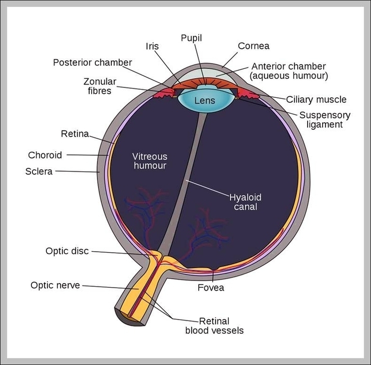
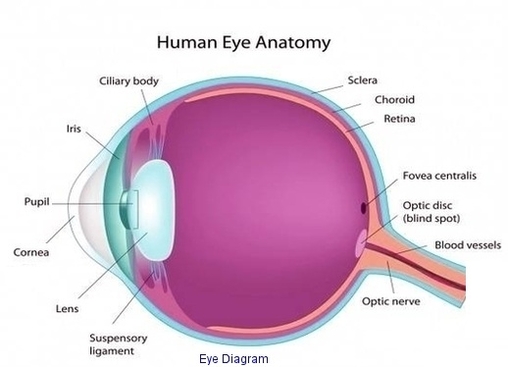
There are many parts of the eye. The anatomy of the eye includes the cornea, pupil, lens, sclera, conjunctiva and more. Parts of the Eye. Muscles in the iris dilate…
There are many parts of the eye. The anatomy of the eye includes the cornea, pupil, lens, sclera, conjunctiva and more. Use this interactive to label different parts of the…
The Anatomy of Human Eye The most complex sensory organs of the human body are the eyes. Every part of the human body is responsible for a specific action, from…
Cow Eye Dissection: Examining Structure and Function Carolina Biological Supply Company1 Introduction The eyes of cows are structurally and functionally similar to the eyes of humans. During this activity, you…
Here are descriptions of some of the main parts of the eye: Cornea: The cornea is the clear outer part of the eye’s focusing system located at the front of…
Human eye, specialized sense organ in humans that is capable of receiving visual images, which are relayed to the brain. The anatomy of the eye includes auxiliary structures, such as…
Eye diagrams usually include voltage and time samples of the data acquired at some sample rate below the data rate. In Figure 1 , the bit sequences 011, 001, 100,…