Posted inDiagrams
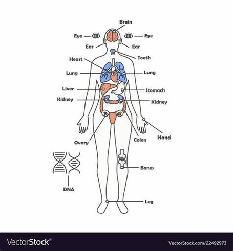
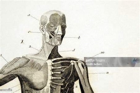
A Skeleton With Labelshuman Skeleton Diagram With Labels Explanation
The human skeleton, an intricate internal framework, provides essential support, protection, and mobility for our bodies. Comprising numerous individual bones and cartilages, it forms the architectural basis upon which our…