Human Heart Anatomy Description Illustration

Human Heart Anatomy Description

The human heart, a muscular organ located between the lungs and slightly to the left of the center, is the main organ of the circulatory system. It is composed of muscle tissue called myocardium, which acts like an engine that pumps blood. The heart is divided into four chambers: two atria (upper chambers) and two ventricles (lower chambers).

The heart’s structure can be likened to a building, with walls, chambers that are like rooms, valves that open and close like doors to the rooms, blood vessels like plumbing pipes that run through a building, and an electrical conduction system like electrical power that runs through a building.

Heart Walls
The heart walls are the muscles that contract (squeeze) and relax to send blood throughout your body. A layer of muscular tissue called the septum divides your heart walls into the left and right sides. Your heart walls have three layers: Endocardium (inner layer), Myocardium (muscular middle layer), and Epicardium (protective outer layer).

Heart Chambers
Your heart has four separate chambers. You have two chambers on the top (atrium, plural atria) and two on the bottom (ventricles), one on each side of your heart. The two atria act as receiving chambers for blood entering the heart; the more muscular ventricles pump the blood out of the heart.

Heart Valves
The heart valves ensure that the blood keeps flowing in the right direction. They act like doors, allowing blood to flow from the atria to the ventricles, and from the ventricles into the two main arteries (the pulmonary artery and the aorta), but not the other way around.

Blood Vessels
The blood vessels, including arteries and veins, connect to the heart, carrying blood throughout the body. The superior vena cava carries venous blood from the head, chest, and arms, while the inferior vena cava carries blood from the abdomen, pelvic region, and legs.

Electrical Conduction System
The heart beats due to electrical impulses. These impulses cause the heart muscle to contract, pumping blood through the heart’s chambers. The brain and nervous system direct your heart’s function.

Function of the Heart
The heart’s main function is to move blood throughout your body. Blood brings oxygen and nutrients to your cells. It also takes away carbon dioxide and other waste so other organs can dispose of them.

In conclusion, the human heart, with its complex structure and intricate functions, serves as the vital engine that powers the circulatory system. Its continuous and coordinated efforts ensure the delivery of oxygen and nutrients to every cell in the body, highlighting its indispensable role in sustaining life..

Human Heart Anatomy Description Illustration Diagram - Human Heart Anatomy Description Illustration Chart - Human anatomy diagrams and charts explained. This anatomy system diagram depicts Human Heart Anatomy Description Illustration with parts and labels. Best diagram to help learn about health, human body and medicine.

Human Heart Anatomy Description Illustration

Human Digestive System Description Illustration

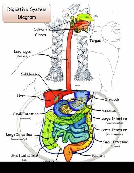
The Human Digestive System

The human digestive system is a complex network of organs that work together to break down food, absorb nutrients, and eliminate waste. It consists of the gastrointestinal (GI) tract and the biliary system.

Gastrointestinal (GI) Tract

The GI tract is a series of hollow organs connected to each other, leading from the mouth to the anus. It includes:

1. Mouth: Digestion begins in the mouth. The salivary glands produce saliva, which mixes with the food to start breaking it down into a form the body can absorb and use.

2. Esophagus: The esophagus is a muscular tube that connects the mouth to the stomach. It uses rhythmic muscle movements (called peristalsis) to force food from the throat into the stomach.

3. Stomach: The stomach secretes acid and enzymes that digest food. The stomach muscles contract periodically, churning food to enhance digestion.

4. Small Intestine: The small intestine is where most of the digestion and absorption of nutrients occur. It is divided into three parts: the duodenum, the jejunum, and the ileum.

5. Large Intestine: The large intestine absorbs water and electrolytes, forming feces. It includes the cecum, ascending colon, transverse colon, descending colon, and sigmoid colon.

6. Anus: The rectum stores feces until they can be eliminated from the body through the anus.

Biliary System

The biliary system includes the liver, gallbladder, pancreas, and bile ducts. These organs produce and secrete bile and enzymes that aid in digestion.

Digestive Process

The digestive process involves the breakdown of food into smaller components until they can be absorbed and assimilated into the body. The body uses these nutrients for energy, growth, and cell repair.

Importance of Digestion

Digestion is crucial because the body needs nutrients from food and liquids to stay healthy and function properly. These nutrients include carbohydrates, proteins, fats, vitamins, minerals, and water.

Digestive Health Conditions

Human Digestive System Description Illustration Diagram - Human Digestive System Description Illustration Chart - Human anatomy diagrams and charts explained. This anatomy system diagram depicts Human Digestive System Description Illustration with parts and labels. Best diagram to help learn about health, human body and medicine.

Human Digestive System Description Illustration

Ear Anatomysimple Ear Anatomy Description

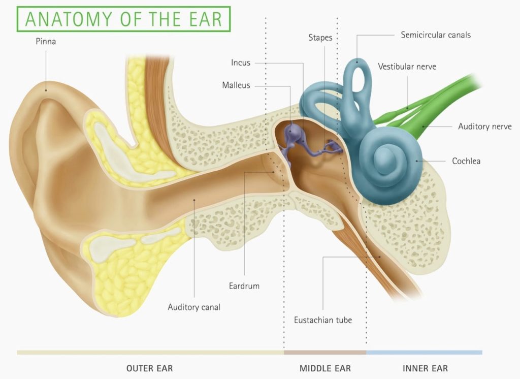
The Anatomy of the Human Ear

The human ear is a complex organ that serves two primary functions: hearing and maintaining balance. It is typically divided into three main parts: the outer ear, the middle ear, and the inner ear.

1. Outer Ear

The outer ear consists of the visible portion called the auricle or pinna, and the short external auditory canal. The auricle, made up of cartilage and skin, comes in various shapes and sizes, contributing to our unique appearance. The function of the outer ear is to collect sound waves and guide them to the tympanic membrane, commonly known as the eardrum.

2. Middle Ear

The middle ear is an air-filled cavity in the temporal bone. It houses three tiny bones — the malleus (hammer), incus (anvil), and stapes (stirrup), collectively known as the auditory ossicles. These bones conduct sound from the tympanic membrane to the inner ear.

3. Inner Ear

The inner ear, also known as the labyrinth, is a complex system of fluid-filled passages and cavities located deep within the temporal bone. It consists of two functional units: the vestibular apparatus, which maintains balance, and the cochlea, which analyzes sound. These sensory organs are highly specialized endings of the eighth cranial nerve, also known as the vestibulocochlear nerve.

Conclusion

The human ear is a marvel of biological engineering, capable of detecting a wide range of sounds and helping us maintain our balance. Its intricate structure and function are a testament to the complexity and sophistication of human anatomy. Understanding the ear’s anatomy not only provides insight into how we hear and maintain balance but also aids in the diagnosis and treatment of related disorders..

Ear Anatomysimple Ear Anatomy Description Diagram - Ear Anatomysimple Ear Anatomy Description Chart - Human anatomy diagrams and charts explained. This anatomy system diagram depicts Ear Anatomysimple Ear Anatomy Description with parts and labels. Best diagram to help learn about health, human body and medicine.

Ear Anatomysimple Ear Anatomy Description

Scottish Organ Donors Illustration Description Example With Labels

Scottish Organ Donors

Organ donation in Scotland is a significant aspect of the country’s healthcare system. It involves the process of giving organs or tissues to help save or improve the lives of others who are critically ill. The system is managed by the NHS Organ Donor Register.

Opt-Out System
cotland operates an opt-out system for organ donation. This means that all adults in Scotland are automatically considered as potential donors unless they choose to opt out. The law changed to this system on March 26, 2021. However, medical staff will always consult the family about the individual’s latest views to ensure donation does not proceed if the person was against it.

Registration

Individuals have the option to register their decision about organ donation. They can choose to donate all or some types of organs and tissues after their death. Alternatively, they can register not to be a donor, which means opting out of the donation of all organs and tissues. It is recommended that individuals discuss their decision with family and friends.

Living Donation

In addition to posthumous organ donation, Scotland also recognizes living donation. This is particularly relevant for kidney donations, as there are currently over 400 people in Scotland in need of a kidney. Living kidney donation is an exceptional gift that can transform the life of someone on the transplant waiting list.

Impact of Donation

Organ donation has a profound impact on the lives of recipients. It not only provides a second chance at life for those who are critically ill but also improves the quality of life for many more. The stories of donor families and recipients highlight the life-changing impact of organ donation.

Conclusion

Organ donation in Scotland is a testament to the power of altruism and medical advancement. It is a personal decision that has far-reaching effects, offering hope and life to those in need. The opt-out system, while respecting individual choice, aims to increase the number of donors and thereby save more lives. As such, organ donation represents a vital part of Scotland’s healthcare landscape.

Scottish Organ Donors Illustration Description Example With Labels Diagram - Scottish Organ Donors Illustration Description Example With Labels Chart - Human anatomy diagrams and charts explained. This anatomy system diagram depicts Scottish Organ Donors Illustration Description Example With Labels with parts and labels. Best diagram to help learn about health, human body and medicine.

Scottish Organ Donors Illustration Description Example With Labels

Abdominal Muscles Description

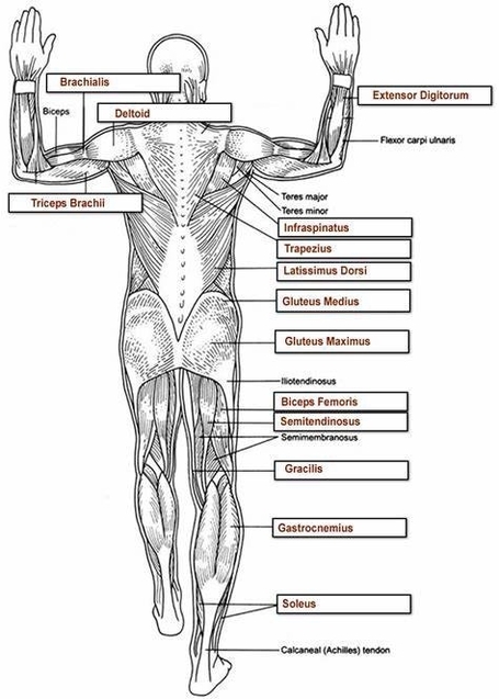
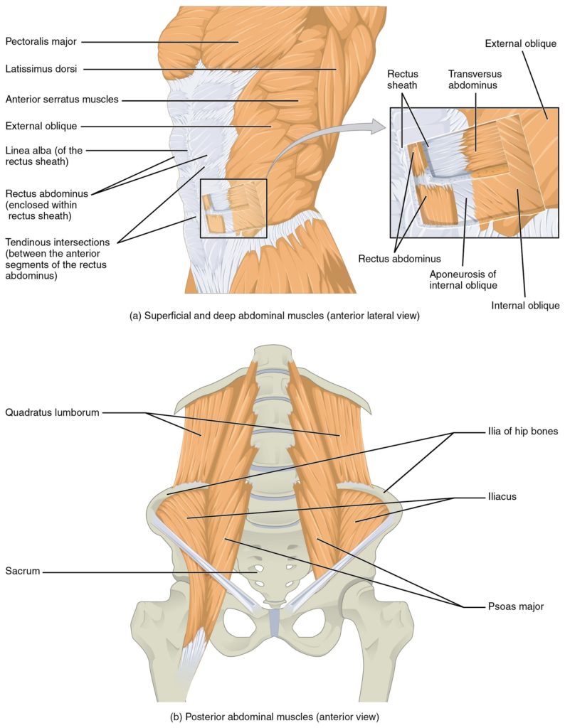
abdominal muscles. These crucial muscles play a pivotal role in our daily lives, from supporting our posture to aiding essential bodily functions. In this concise exploration, I’ll provide an overview of their anatomy, functions, and significance.

## Abdominal Muscles: Anatomy and Function

### Overview
Your abdominal muscles form a robust network of bands that line the walls of your abdomen, which is essentially the trunk of your body. Positioned toward the front, between your ribs and pelvis, these muscles contribute significantly to your overall well-being.

### The Five Main Abdominal Muscles
1. External Obliques:
– These muscles are like sturdy girders flanking the rectus abdominis (more on that shortly).
– Situated on each side of the abdomen, they run from the sides of your body toward the middle.
– Their primary function is to allow trunk rotation, enabling you to twist from side to side.

2. Internal Obliques:
– Nestled just inside your hip bones, the internal obliques mirror the external obliques.
– Like their external counterparts, they also run from the sides of your trunk toward the middle.
– Together, the internal and external obliques form a dynamic duo, supporting lateral movements.

3. Pyramidalis:
– This small, triangular muscle resides low in your pelvis.
– Its role is to help maintain internal pressure within your abdomen.
– While less prominent than other abdominal muscles, it contributes to overall stability.

4. Rectus Abdominis:
– The rectus abdominis muscles are the showstoppers—the ones that often evoke the coveted “six-pack” appearance.
– These paired muscles extend down the middle of your abdomen, spanning from your ribs to the front of your pelvis.
– Their critical functions include holding your internal organs in place and stabilizing your body during movement.

5. Transversus Abdominis:
– The deepest of the abdominal muscles, the transversus abdominis wraps around your abdomen like a supportive corset.
– It plays a vital role in maintaining consistent internal pressure within the abdominal cavity.
– Additionally, it provides core support and contributes to spinal stability.

### Functions of Abdominal Muscles
1. Essential Bodily Functions:
– Your abdominal muscles are unsung heroes in everyday activities.
– They assist with urination, defecation, coughing, sneezing, and even vomiting.
– During childbirth, they increase intra-abdominal pressure, facilitating the birthing process.

2. Organ Support and Protection:
– These muscles hold your internal organs in place, safeguarding vital structures such as the stomach, intestines, pancreas, liver, and gallbladder.
– Imagine them as a protective shield for your precious innards.

3. Posture Maintenance:
– Abdominal muscles contribute significantly to maintaining an upright posture.
– Whether you’re sitting, standing, or bending over, they work tirelessly to keep you balanced.

4. Core Stability:
– The deep abdominal muscles, including the transversus abdominis, form part of your core.
– A stable core is essential for overall body stability, balance, and efficient movement.

5. Spinal Support:
– When you lift, twist, or engage in physical activities, your abdominal muscles collaborate with your back muscles to support your spine.
– They play a crucial role in preventing strain and injury.

In summary, these abdominal muscles are more than mere aesthetics—they’re the unsung champions of our daily lives. So, the next time you laugh, bend, or sing, remember to thank your abdominal muscles for their tireless efforts! ????


I’ve provided a concise yet informative overview of abdominal muscles, emphasizing their functions and significance. If you’d like further details or have any specific questions, feel free to ask! ??

: [Cleveland Clinic – Abdominal Muscles: Anatomy and Function](https://my.clevelandclinic.org/health/body/21755-abdominal-muscles).
ou

Abdominal Muscles Description Diagram - Abdominal Muscles Description Chart - Human anatomy diagrams and charts explained. This anatomy system diagram depicts Abdominal Muscles Description with parts and labels. Best diagram to help learn about health, human body and medicine.

Abdominal Muscles Description

Rn Case Manager Job Description Image

Part One What is an RN Case Manager? A case manager is a specialized Registered Nurse (RN) that works with patients and providers to determine the specific care that is required and the best options for that care. Through a collaboration with multiple specialties, case managers ensure the patient is receiving quality medical care.
This Case Manager job description template is optimized for posting on online job boards or careers pages. It is easy to customize as a nurse, medical or mental health case worker for your company. Coordinating and providing care that is safe, timely, effective, efficient, equitable, and client-centered

Rn Case Manager Job Description Image Diagram - Rn Case Manager Job Description Image Chart - Human anatomy diagrams and charts explained. This anatomy system diagram depicts Rn Case Manager Job Description Image with parts and labels. Best diagram to help learn about health, human body and medicine.

Rn Case Manager Job Description Image