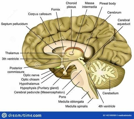
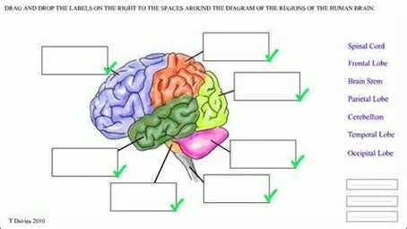
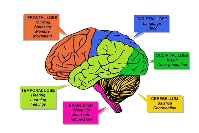
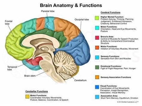
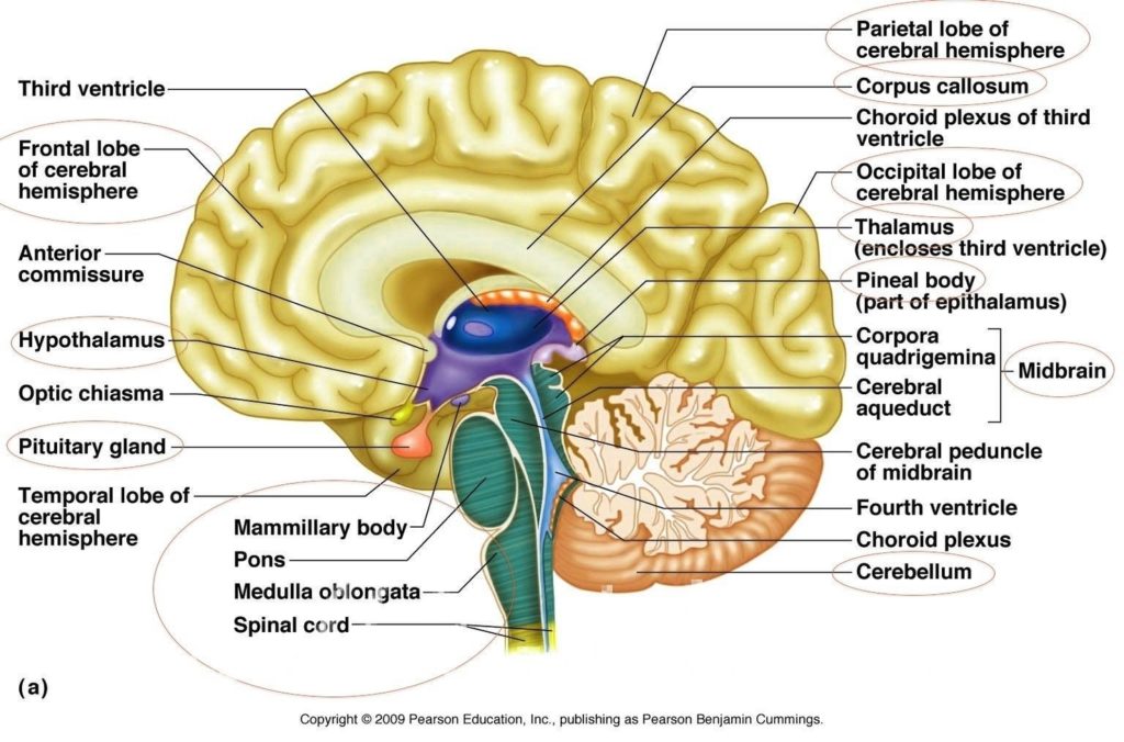
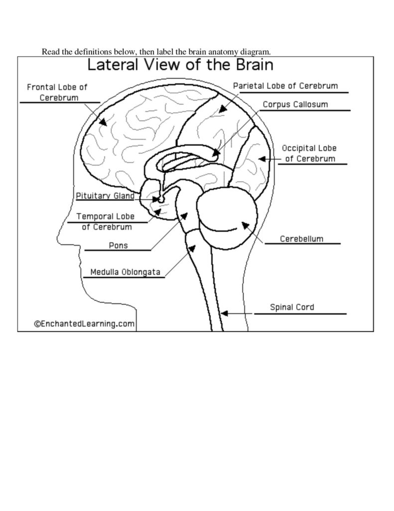
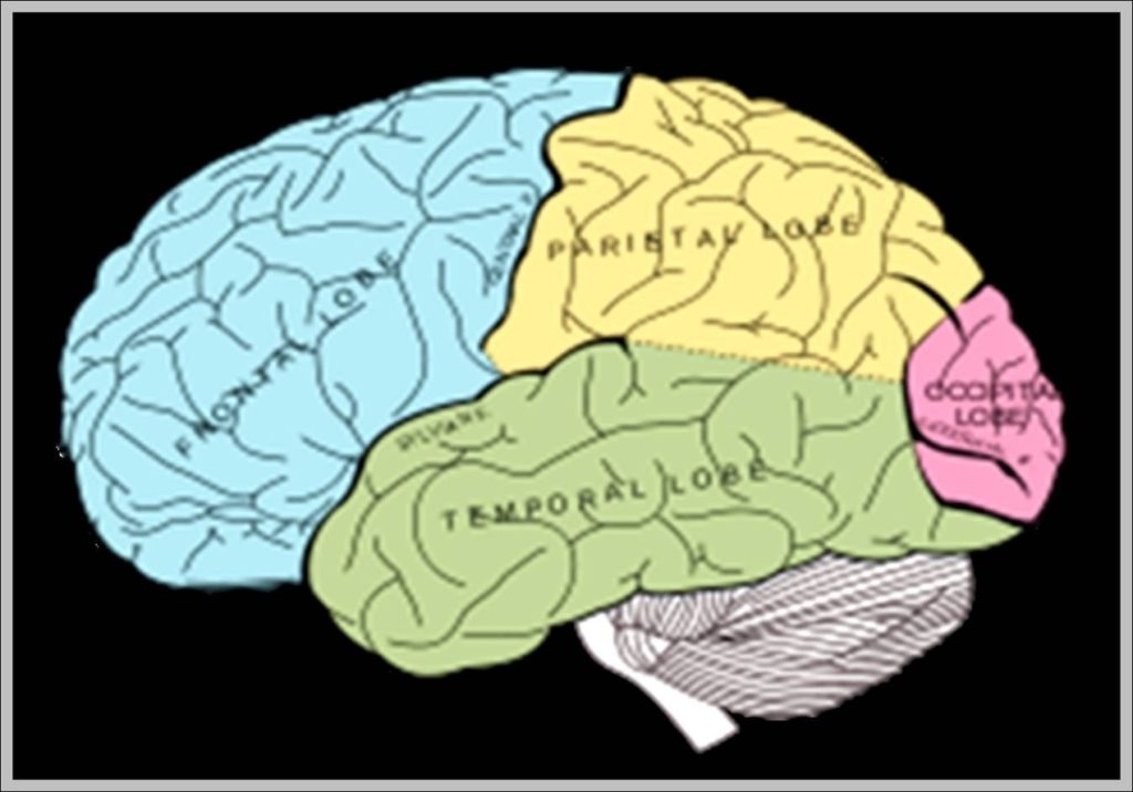
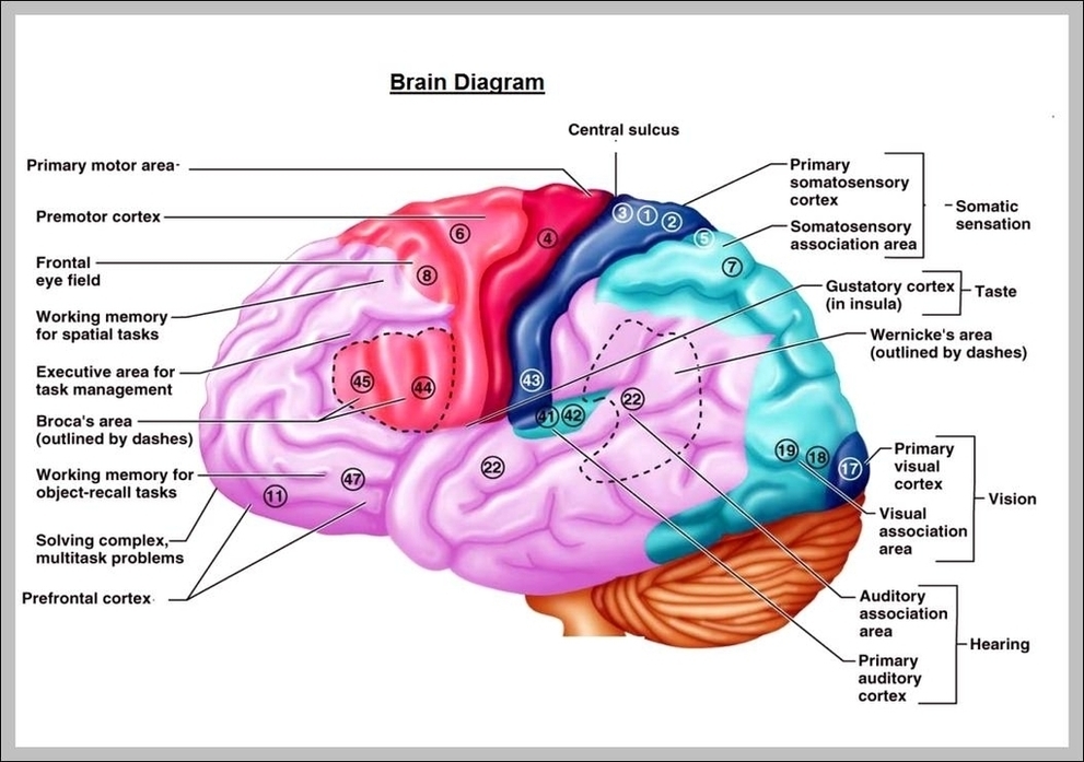
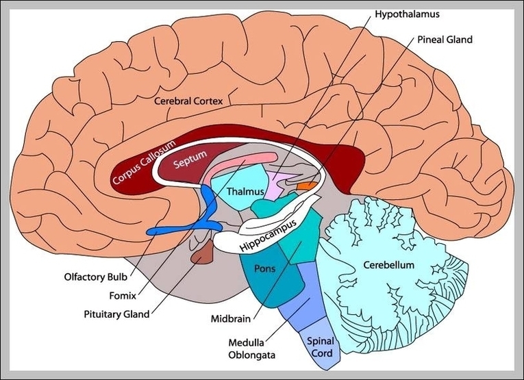
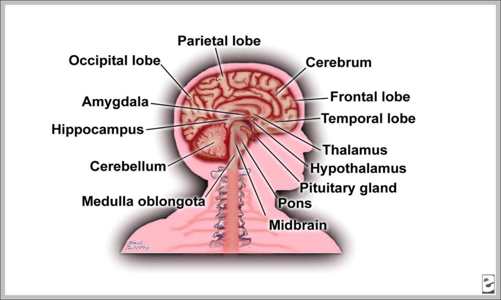
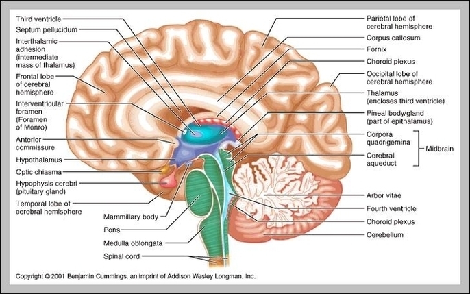
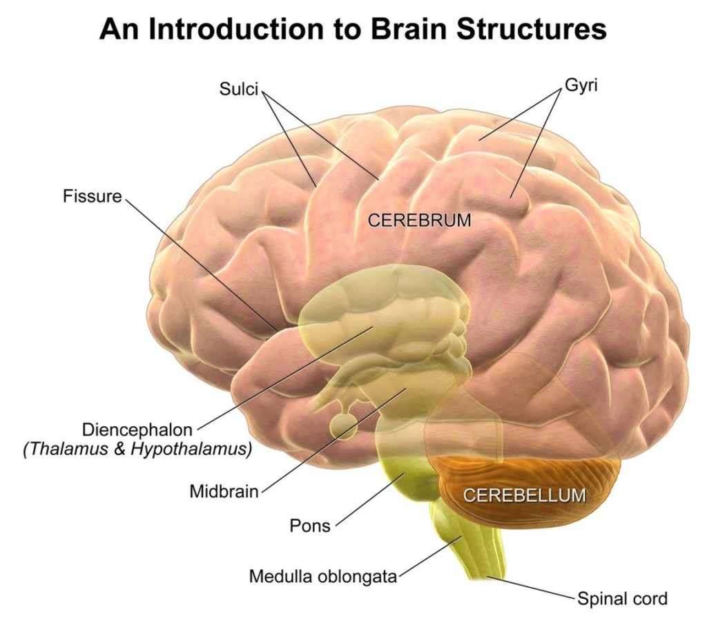
Human Brain Anatomy Examined

The Human Brain: An Examination of Its Anatomy

The human brain, a complex organ, is the central component of the nervous system. It controls thought, memory, emotion, touch, motor skills, vision, breathing, temperature, hunger, and every process that regulates our body.

Composition

Weighing about 3 pounds in the average adult, the brain is about 60% fat. The remaining 40% is a combination of water, protein, carbohydrates, and salts. The brain is primarily composed of nerve cells, also called neurons. It contains blood vessels and nerves, including neurons and glial cells.

Gray Matter and White Matter

Gray and white matter are two different regions of the central nervous system. In the brain, gray matter refers to the darker, outer portion, while white matter describes the lighter, inner section underneath. Gray matter is primarily composed of neuron somas (the round central cell bodies), and white matter is mostly made of axons (the long stems that connect neurons together) wrapped in myelin (a protective coating). Gray matter is primarily responsible for processing and interpreting information, while white matter transmits that information to other parts of the nervous system.

Main Parts of the Brain

At a high level, the brain can be divided into the cerebrum, brainstem, and cerebellum.

1. Cerebrum: The cerebrum (front of the brain) comprises gray matter (the cerebral cortex) and white matter at its center. The largest part of the brain, the cerebrum initiates and coordinates movement and regulates temperature. Other areas of the cerebrum enable speech, judgment, thinking and reasoning, problem-solving, emotions, and learning. Other functions relate to vision, hearing, touch, and other senses.

2. Cerebral Cortex: The cortex has a large surface area due to its folds, and comprises about half of the brain’s weight. The cerebral cortex is divided into two halves, or hemispheres. It is covered with ridges (gyri) and folds (sulci).

3. Brainstem: The brainstem consists of the midbrain, the pons, and the medulla oblongata.

4. Cerebellum: The cerebellum is connected to the brainstem by three pairs of nerve tracts called cerebellar peduncles.

Functioning

The brain sends and receives chemical and electrical signals throughout the body. Different signals control different processes, and your brain interprets each. Some make you feel tired, for example, while others make you feel pain. Some messages are kept within the brain, while others are relayed through the spine and across the body’s vast network of nerves to distant extremities.

Conclusion

The human brain, with its intricate structure and complex functioning, is a marvel of nature. Its anatomy and physiology are key to our understanding of ourselves and the world around us. As we continue to explore its mysteries, we gain deeper insights into the workings of this remarkable organ..

Human Brain Anatomy Examined Diagram - Human Brain Anatomy Examined Chart - Human anatomy diagrams and charts explained. This anatomy system diagram depicts Human Brain Anatomy Examined with parts and labels. Best diagram to help learn about health, human body and medicine.

Human Brain Anatomy Examined

Human Brain Diagram Blank

The Human Brain

The human brain, the central organ of the human nervous system, is a complex structure that controls most of the body’s activities. It processes, integrates, and coordinates information received from the sense organs, and makes decisions as to the instructions sent to the rest of the body.

Composition

Weighing about 3 pounds in the average adult, the brain is about 60% fat. The remaining 40% is a combination of water, protein, carbohydrates, and salts. The brain is not a muscle but contains blood vessels and nerves, including neurons and glial cells.
tructure

The brain consists of the cerebrum, the brainstem, and the cerebellum. The cerebrum, the largest part of the human brain, consists of two cerebral hemispheres. Each hemisphere has an inner core composed of white matter, and an outer surface – the cerebral cortex – composed of grey matter.

Cerebral Cortex

The cortex has an outer layer, the neocortex, and an inner allocortex. The neocortex is made up of six neuronal layers, while the allocortex has three or four. Each hemisphere is conventionally divided into four lobes – the frontal, temporal, parietal, and occipital lobes.

Functions

The frontal lobe is associated with executive functions including self-control, planning, reasoning, and abstract thought, while the occipital lobe is dedicated to vision. Within each lobe, cortical areas are associated with specific functions, such as the sensory, motor, and association regions.

Hemispheres

Although the left and right hemispheres are broadly similar in shape and function, some functions are associated with one side, such as language in the left and visual-spatial ability in the right. The hemispheres are connected by commissural nerve tracts, the largest being the corpus callosum.

Brainstem and Cerebellum

The cerebrum is connected by the brainstem to the spinal cord. The brainstem consists of the midbrain, the pons, and the medulla oblongata. The cerebellum is connected to the brainstem by three pairs of nerve tracts called cerebellar peduncles.

Other Structures

Underneath the cerebral cortex are several important structures, including the thalamus, the epithalamus, the pineal gland, the hypothalamus, the pituitary gland, and the subthalamus; the limbic structures, including the amygdalae and the hippocampi, the claustrum, the various nuclei of the basal ganglia, the basal forebrain structures, and the three circumventricular organs.

Conclusion

The human brain, with its intricate structure and complex functions, is a marvel of nature. It is the command center for the human nervous system, receiving signals from the body’s sensory organs and outputting information to the muscles. Its study continues to be a fascinating field, offering insights into our behavior, cognition, and the very essence of what makes us human..

Human Brain Diagram Blank Diagram - Human Brain Diagram Blank Chart - Human anatomy diagrams and charts explained. This anatomy system diagram depicts Human Brain Diagram Blank with parts and labels. Best diagram to help learn about health, human body and medicine.

Human Brain Diagram Blank

Human Brain External Structure

The human brain, the main organ of the central nervous system, is a complex structure that controls thought, memory, emotion, touch, motor skills, vision, breathing, temperature, hunger, and every process that regulates our body. It is located in the head, protected by the cranium?.

The brain is made up of several distinct parts, each responsible for different functions. The largest part is the cerebrum, which is responsible for sensory interpretation, thought processing, and voluntary muscle activity. The cerebrum consists of two cerebral hemispheres, each having an inner core composed of white matter, and an outer surface – the cerebral cortex – composed of grey matter. The cortex has an outer layer, the neocortex, and an inner allocortex.

The cerebral hemispheres in humans have many folds to increase the surface area of the brain. The ridges are called gyri and the grooves are called sulci. Large sulci are often called fissures. The cerebrum is organized into folds called gyri and grooves called sulci. The cerebellum sits behind (posterior) and below (inferior) the cerebrum. The brainstem connects the brain with the spinal cord and exits from the ventral side of the brain.

The cerebral hemispheres of the brain are divided into four lobes. The frontal lobes are the most rostral, located in the front of the brain and are responsible for higher-level executive functions, like attention, critical thinking, and impulse control. The frontal lobes are also the location of the primary motor cortex, the region of the brain responsible for planning and executing movement.

The central sulcus lies caudal to the frontal lobe and divides the frontal lobes from the parietal lobes. The parietal lobes are important for processing sensory information. The primary somatosensory cortex is located in the postcentral gyrus of the parietal lobe and is responsible for the perception of touch and pain.

Three layers of protective covering called meninges surround the brain and the spinal cord. The outermost layer, the dura mater, is thick and tough. It includes two layers: The periosteal layer of the dura mater lines the inner dome of the skull (cranium) and the meningeal layer is below that.

The brain sends and receives chemical and electrical signals throughout the body. Different signals control different processes, and your brain interprets each. Some make you feel tired, for example, while others make you feel pain. Some messages are kept within the brain, while others are relayed through the spine and across the body’s vast network

Human Brain External Structure Diagram - Human Brain External Structure Chart - Human anatomy diagrams and charts explained. This anatomy system diagram depicts Human Brain External Structure with parts and labels. Best diagram to help learn about health, human body and medicine.

Human Brain External Structure

Inner Ear And Brain

The Inner Ear and Its Connection to the Brain

The inner ear, also known as the labyrinth, is a complex structure that plays a crucial role in our ability to hear and maintain balance. It is the deepest part of the ear, located in a small cavity within the skull bones on both sides of the head.

Anatomy of the Inner Ear

The inner ear consists of three main parts:

1. Cochlea: This is the auditory area of the inner ear that changes sound waves into nerve signals. It is shaped like a snail’s shell and filled with fluid. Inside the cochlea is a sensitive structure called the organ of Corti, which contains tiny hairs that pick up the vibrations from sound waves.

2. Semicircular Canals: These canals sense balance and posture to assist in equilibrium. They are filled with fluid and lined with fine hairs, similar to the cochlea, but these hairs pick up body movements instead of sounds.

3. Vestibule: This is the area of the inner ear cavity that lies between the cochlea and semicircular canals, also assisting in equilibrium.

Function of the Inner Ear

The inner ear has two main functions: hearing and balance.

*Hearing*: The cochlea works with parts of the outer and middle ear to help us hear sounds. Sound waves travel from the outer ear to the eardrum in the middle ear, causing it to vibrate. This vibration moves the three tiny bones in the middle ear, leading to pressure waves that make the fluid inside the cochlea move. The movement of fluid in the inner ear makes the tiny hairs in the cochlea bend and move. These “dancing” hairs convert the movement from sound waves into electrical signals.

*Balance*: The balance parts of the inner ear are the vestibule and the semicircular canals. The semicircular canals sit at right angles to each other, which helps them measure motions no matter what position you’re in. When your head moves around, the fluid inside the semicircular canals shifts around.

Connection to the Brain

The electrical signals generated by the inner ear are sent to the brain through the hearing (auditory) nerves. The brain then interprets these signals, allowing us to hear and understand sounds. At the same time, the inner ear monitors our movements, alerting the brain to changes so it can let our body know what to do to stay balanced.

Conclusion

The inner ear is a remarkable structure that not only enables us to hear the world around us but also helps us maintain our balance. Its intricate design and function illustrate the complexity of the human body and the importance of maintaining ear health. Problems with the inner ear can result in hearing loss and balance issues, highlighting the critical role it plays in our daily lives..

Inner Ear And Brain Diagram - Inner Ear And Brain Chart - Human anatomy diagrams and charts explained. This anatomy system diagram depicts Inner Ear And Brain with parts and labels. Best diagram to help learn about health, human body and medicine.

Inner Ear And Brain

Human Brain Partsbrain Parts Anatomy

The human brain, an organ of immense complexity and the center of our consciousness, is divided into several distinct parts, each with its own specific functions.

1. Cerebrum
The cerebrum, the largest part of the brain, initiates and coordinates movement and regulates temperature. It is responsible for higher functions like speech, judgment, thinking, reasoning, problem-solving, emotions, and learning. The cerebrum is divided into two hemispheres, each with four sections or lobes: frontal, parietal, temporal, and occipital.

– Frontal Lobe: This lobe is associated with reasoning, motor skills, higher level cognition, and expressive language.
– Parietal Lobe: The parietal lobe processes tactile sensory information such as pressure, touch, and pain.
– Temporal Lobe: This lobe interprets sounds and language we hear. It is also heavily associated with the formation of memories.
– Occipital Lobe: The occipital lobe is primarily responsible for vision.

2. Cerebral Cortex
The cerebral cortex, the outermost layer of the cerebrum, is responsible for consciousness, higher-order thinking, imagination, information processing, language, memory, perception, reasoning, sensation, and voluntary physical action.

3. Cerebellum
The cerebellum, located at the back of the brain, is responsible for coordinating voluntary movements, maintaining posture, and balancing.

4. Brainstem
The brainstem controls automatic functions such as breathing, heart rate, and blood pressure.

5. Limbic System
The limbic system, located deep within the brain, is responsible for emotions, survival instincts, and memory.

6. Gray and White Matter
The brain is composed of gray and white matter. Gray matter, found in the outer portion of the brain, is primarily composed of neuron somas (the round central cell bodies). White matter, located in the inner section, is mostly made of axons (the long stems that connect neurons together) wrapped in myelin (a protective coating). Gray matter is primarily responsible for processing and interpreting information, while white matter transmits that information to other parts of the nervous system.

In conclusion, the human brain is a marvel of biological engineering, with each part playing a crucial role in maintaining our daily functions and experiences. Despite the vast knowledge we have gained about the brain, there is still much to learn about this complex organ..

Human Brain Partsbrain Parts Anatomy Diagram - Human Brain Partsbrain Parts Anatomy Chart - Human anatomy diagrams and charts explained. This anatomy system diagram depicts Human Brain Partsbrain Parts Anatomy with parts and labels. Best diagram to help learn about health, human body and medicine.

Human Brain Partsbrain Parts Anatomy

Brain Diagram Worksheet

A brain worksheet is an educational tool designed to help individuals learn about the structure and functions of the human brain. It often includes diagrams, labeling exercises, quizzes, and other interactive elements to facilitate learning.
tructure of the Brain

Brain worksheets typically focus on the major parts of the brain, including the cerebrum, cerebellum, and brainstem. Each of these parts is further divided into various regions, each with its specific functions.

1. Cerebrum: The largest part of the brain, responsible for higher brain functions such as thinking, learning, and consciousness. It’s divided into two hemispheres, each consisting of four lobes: frontal, parietal, occipital, and temporal.

2. Cerebellum: Located at the back of the brain, it’s responsible for coordinating voluntary movements, balance, and muscle coordination.

3. Brainstem: It connects the cerebrum and cerebellum to the spinal cord and controls automatic functions necessary for survival, like heart rate, breathing, and blood pressure.

Labeling Exercises

Labeling exercises are a common feature of brain worksheets. They involve diagrams of the brain with parts marked for students to identify and label. This helps reinforce the understanding of brain anatomy and the location of different structures.

Quizzes and Interactive Elements

Brain worksheets often include quizzes and other interactive elements to test knowledge and reinforce learning. These can range from multiple-choice questions about brain functions to matching exercises where students link brain parts to their functions.

Psychoeducation
ome brain worksheets are used in psychoeducation, helping individuals understand how the brain works in relation to mental health. For example, they may explain how the brain changes during the fight-or-flight response, anger, or drug intoxication.

Conclusion

In summary, brain worksheets are valuable tools for learning about the complex structure and functions of the human brain. They use a variety of methods, including diagrams, labeling exercises, and quizzes, to make the learning process interactive and engaging. Whether used in a classroom, therapy setting, or for self-study, brain worksheets can significantly enhance understanding of this vital organ..

Brain Diagram Worksheet Diagram - Brain Diagram Worksheet Chart - Human anatomy diagrams and charts explained. This anatomy system diagram depicts Brain Diagram Worksheet with parts and labels. Best diagram to help learn about health, human body and medicine.

Brain Diagram Worksheet

Parts And Functions Of The Brain

The human brain, the most complex organ in the body, is responsible for controlling thought, memory, emotion, touch, motor skills, vision, breathing, temperature, hunger, and every process that regulates our body. It consists of billions of neurons (nerve cells) that communicate through intricate networks?. The brain can be divided into four major regions: the cerebrum, the cerebellum, the brainstem, and the diencephalon.

1. Cerebrum
The cerebrum, the largest part of the brain, initiates and coordinates movement and regulates temperature. It comprises gray matter (the cerebral cortex) and white matter at its center. The cerebral cortex is responsible for higher-order functions such as consciousness, thinking, imagination, language, memory, perception, reasoning, sensation, and voluntary physical action. It is divided into four lobes, each associated with different functions:

– Frontal Lobe: Associated with reasoning, motor skills, higher-level cognition, and expressive language. Damage to this lobe can lead to changes in sexual habits, socialization, attention, and increased risk-taking.
– Parietal Lobe: Processes tactile sensory information such as pressure, touch, and pain.
– Temporal Lobe: Responsible for interpreting sounds and language. It also plays a significant role in the formation of memories.
– Occipital Lobe: Primarily associated with interpreting visual stimuli and information.

2. Cerebellum
The cerebellum coordinates voluntary movements such as posture, balance, coordination, and speech, resulting in smooth and balanced muscular activity.

3. Brainstem
The brainstem controls the flow of messages between the brain and the rest of the body, and it also controls basic body functions such as breathing, swallowing, heart rate, blood pressure, consciousness, and whether one is awake or sleepy.

4. Diencephalon
The diencephalon contains structures such as the thalamus and hypothalamus. The thalamus acts as a kind of relay station for signals coming into the brain, while the hypothalamus is responsible for maintaining your body’s internal balance by regulating various bodily functions such as heart rate and body temperature.

In conclusion, the brain is a complex organ that performs a multitude of functions that are essential for human life. It is responsible for everything we think, feel, and do. Understanding the brain’s structure and functions helps us understand ourselves better and provides insights into how we interact with the world around us..

Parts And Functions Of The Brain Diagram - Parts And Functions Of The Brain Chart - Human anatomy diagrams and charts explained. This anatomy system diagram depicts Parts And Functions Of The Brain with parts and labels. Best diagram to help learn about health, human body and medicine.

Parts And Functions Of The Brain

Parts Of The Brain Diagram And Function

The human brain, the most complex organ in the body, is responsible for controlling thought, memory, emotion, touch, motor skills, vision, breathing, temperature, hunger, and every process that regulates our body. It is made up of billions of neurons and has several specialized parts, each involved in important functions.

1. Cerebrum
The cerebrum, the largest part of the brain, initiates and coordinates movement and regulates temperature. It comprises gray matter (the cerebral cortex) and white matter at its center. The cerebral cortex is responsible for higher-order functions such as consciousness, imagination, information processing, language, memory, perception, reasoning, sensation, and voluntary physical action. It is divided into four lobes:

– Frontal Lobe: Associated with reasoning, motor skills, higher-level cognition, and expressive language. Damage to this lobe can lead to changes in sexual habits, socialization, attention, and increased risk-taking.
– Parietal Lobe: Processes tactile sensory information such as pressure, touch, and pain.
– Occipital Lobe: Responsible for vision.
– Temporal Lobe: Interprets sounds and language we hear. It also heavily associated with the formation of memories.

2. Cerebellum
The cerebellum coordinates balance and muscle activity. It plays a vital role in motor control and is also involved in some cognitive functions such as attention and language, and in regulating fear and pleasure responses.

3. Brainstem
The brainstem controls basic functions like breathing. It acts as a relay center connecting the cerebrum and cerebellum to the spinal cord, performing many automatic functions such as breathing, heart rate, body temperature, wake and sleep cycles, digestion, sneezing, and coughing.

4. Diencephalon
The diencephalon regulates processes like sleep and body temperature. It consists of structures such as the thalamus and hypothalamus which are responsible for such functions as motor control, relaying sensory information, and controlling autonomic functions.

5. Limbic System
The limbic system, often referred to as the “emotional brain”, is involved in many of our emotions and motivations, particularly those related to survival such as fear and anger.

6. Gray and White Matter
Gray matter is primarily composed of neuron somas (the round central cell bodies), and white matter is mostly made of axons (the long stems that connect neurons together) wrapped in myelin (a protective coating). Gray matter is primarily responsible for processing and interpreting information, while white matter transmits that information to other parts of the nervous system.

In conclusion, the brain is a complex organ with various parts working together to regulate and control bodily functions. Each part plays a unique role, and understanding these parts can help us understand how disease and damage may affect the brain and its ability to function..

Parts Of The Brain Diagram And Function Diagram - Parts Of The Brain Diagram And Function Chart - Human anatomy diagrams and charts explained. This anatomy system diagram depicts Parts Of The Brain Diagram And Function with parts and labels. Best diagram to help learn about health, human body and medicine.

Parts Of The Brain Diagram And Function

Brain Anatomy And Physiology Definition

Brain Anatomy and Physiology

The brain, a complex organ, is the central hub of the nervous system, controlling thought, memory, emotion, touch, motor skills, vision, breathing, temperature, hunger, and every process that regulates our body. It integrates sensory information and directs motor responses.

Composition

Weighing about 3 pounds in the average adult, the brain is about 60% fat. The remaining 40% is a combination of water, protein, carbohydrates, and salts. It contains blood vessels and nerves, including neurons and glial cells.

Gray Matter and White Matter

Gray and white matter are two different regions of the central nervous system. In the brain, gray matter refers to the darker, outer portion, while white matter describes the lighter, inner section underneath. Gray matter is primarily composed of neuron somas (the round central cell bodies), and white matter is mostly made of axons (the long stems that connect neurons together) wrapped in myelin (a protective coating). Gray matter is primarily responsible for processing and interpreting information, while white matter transmits that information to other parts of the nervous system.

Functioning

The brain sends and receives chemical and electrical signals throughout the body. Different signals control different processes, and your brain interprets each. Some make you feel tired, for example, while others make you feel pain. Some messages are kept within the brain, while others are relayed through the spine and across the body’s vast network of nerves to distant extremities.

Main Parts of the Brain

At a high level, the brain can be divided into the cerebrum, brainstem, and cerebellum.

– Cerebrum: The cerebrum (front of brain) comprises gray matter (the cerebral cortex) and white matter at its center. The largest part of the brain, the cerebrum initiates and coordinates movement and regulates temperature. Other areas of the cerebrum enable speech, judgment, thinking and reasoning, problem-solving, emotions, and learning.

– Cerebral Cortex: The cortex has a large surface area due to its folds, and comprises about half of the brain’s weight. The cerebral cortex is divided into two halves, or hemispheres. It is covered with ridges (gyri) and folds (sulci).

– Brainstem and Cerebellum: The brainstem and cerebellum control both voluntary movements, such as those involved in walking and in speech, and involuntary movements, such as breathing and reflex actions.

In conclusion, the brain is a marvel of nature, a complex organ that not only controls our bodily functions but also makes us who we are, shaping our thoughts, emotions, and behaviors..

Brain Anatomy And Physiology Definition Diagram - Brain Anatomy And Physiology Definition Chart - Human anatomy diagrams and charts explained. This anatomy system diagram depicts Brain Anatomy And Physiology Definition with parts and labels. Best diagram to help learn about health, human body and medicine.

Brain Anatomy And Physiology Definition

Brain Anatomy Diagram

Brain Anatomy

The brain, a complex organ, controls thought, memory, emotion, touch, motor skills, vision, breathing, temperature, hunger, and every process that regulates our body. It is primarily composed of nerve cells, also known as neurons. Blood vessels supply oxygen and nutrients to the neurons of the brain.

Composition

Weighing about 3 pounds in the average adult, the brain is about 60% fat. The remaining 40% is a combination of water, protein, carbohydrates, and salts. The brain itself is not a muscle. It contains blood vessels and nerves, including neurons and glial cells.

Gray Matter and White Matter

Gray and white matter are two different regions of the central nervous system. In the brain, gray matter refers to the darker, outer portion, while white matter describes the lighter, inner section underneath. Gray matter is primarily composed of neuron somas (the round central cell bodies), and white matter is mostly made of axons (the long stems that connect neurons together) wrapped in myelin (a protective coating). Gray matter is primarily responsible for processing and interpreting information, while white matter transmits that information to other parts of the nervous system.

Brain Function

The brain sends and receives chemical and electrical signals throughout the body. Different signals control different processes, and your brain interprets each. Some make you feel tired, for example, while others make you feel pain. Some messages are kept within the brain, while others are relayed through the spine and across the body’s vast network of nerves to distant extremities.

Main Parts of the Brain and Their Functions

At a high level, the brain can be divided into the cerebrum, brainstem, and cerebellum.

1. Cerebrum: The cerebrum (front of brain) comprises gray matter (the cerebral cortex) and white matter at its center. The largest part of the brain, the cerebrum initiates and coordinates movement and regulates temperature. Other areas of the cerebrum enable speech, judgment, thinking and reasoning, problem-solving, emotions, and learning. Other functions relate to vision, hearing, touch, and other senses.

2. Cerebral Cortex: The cortex has a large surface area due to its folds, and comprises about half of the brain’s weight. The cerebral cortex is divided into two halves, or hemispheres. It is covered with ridges (gyri) and folds (sulci).

3. Brain Stem: The brain stem controls the flow of messages between the brain and the rest of the body, and it also controls basic body functions such as breathing, swallowing, heart rate, blood pressure, consciousness, and whether one is awake or sleepy.

4. Cerebellum: The cerebellum is involved in the coordination of voluntary motor movement, balance and equilibrium and muscle tone.

5. Limbic System: The limbic system is a complex set of structures that lies on both sides of the thalamus, just under the cerebrum. It includes the hypothalamus, the hippocampus, the amygdala, and several other nearby areas. It appears to be primarily responsible for our emotional life, and has a lot to do with the formation of memories.

In conclusion, the brain is a complex organ with various parts working together to regulate our body’s functions and processes. Understanding its anatomy helps us appreciate its importance and the role it plays in our daily lives..

Brain Anatomy Diagram Diagram - Brain Anatomy Diagram Chart - Human anatomy diagrams and charts explained. This anatomy system diagram depicts Brain Anatomy Diagram with parts and labels. Best diagram to help learn about health, human body and medicine.

Brain Anatomy Diagram

Human Brain Anatomy Graphic

Human Brain Anatomy

The human brain, a complex organ, is the central component of the nervous system. It controls thought, memory, emotion, touch, motor skills, vision, breathing, temperature, hunger, and every process that regulates our body.

Composition

Weighing about 3 pounds in the average adult, the brain is about 60% fat. The remaining 40% is a combination of water, protein, carbohydrates, and salts. The brain is primarily composed of nerve cells, also known as neurons, and supportive glial cells. It contains blood vessels and nerves, including neurons and glial cells.

Gray Matter and White Matter

Gray and white matter are two different regions of the central nervous system. In the brain, gray matter refers to the darker, outer portion, while white matter describes the lighter, inner section underneath. Gray matter is primarily composed of neuron somas (the round central cell bodies), and white matter is mostly made of axons (the long stems that connect neurons together) wrapped in myelin (a protective coating). Gray matter is primarily responsible for processing and interpreting information, while white matter transmits that information to other parts of the nervous system.

Main Parts of the Brain

The brain can be divided into the cerebrum, brainstem, and cerebellum.

1. Cerebrum: The cerebrum (front of the brain) comprises gray matter (the cerebral cortex) and white matter at its center. The largest part of the brain, the cerebrum initiates and coordinates movement and regulates temperature. Other areas of the cerebrum enable speech, judgment, thinking and reasoning, problem-solving, emotions, and learning. Other functions relate to vision, hearing, touch, and other senses.

2. Cerebral Cortex: The cortex has a large surface area due to its folds, and comprises about half of the brain’s weight. The cerebral cortex is divided into two halves, or hemispheres. It is covered with ridges (gyri) and folds (sulci).

3. Brainstem: The brainstem consists of the midbrain, the pons, and the medulla oblongata.

4. Cerebellum: The cerebellum is connected to the brainstem by three pairs of nerve tracts called cerebellar peduncles.

Functioning

The brain sends and receives chemical and electrical signals throughout the body. Different signals control different processes, and your brain interprets each. Some make you feel tired, for example, while others make you feel pain. Some messages are kept within the brain, while others are relayed through the spine and across the body’s vast network of nerves to distant extremities. To do this, the central nervous system relies on billions of neurons (nerve cells)..

Human Brain Anatomy Graphic Diagram - Human Brain Anatomy Graphic Chart - Human anatomy diagrams and charts explained. This anatomy system diagram depicts Human Brain Anatomy Graphic with parts and labels. Best diagram to help learn about health, human body and medicine.

Human Brain Anatomy Graphic

Human Brain Diagram Labeled Image

Before we have a look at the brain diagram, it is important to go through a few facts about the brain and its function. This will help you understand the anatomy of the brain better. The average dimension of the adult human brain is 5.5 inches in width and 6.5 inches in length.
The diagram of the brain is useful for both Class 10 and 12. It is one among the few topics having the highest weightage of marks and is frequently asked in the examinations. A well-labelled diagram of a human brain is given below for further reference.

Human Brain Diagram Labeled Image Diagram - Human Brain Diagram Labeled Image Chart - Human anatomy diagrams and charts explained. This anatomy system diagram depicts Human Brain Diagram Labeled Image with parts and labels. Best diagram to help learn about health, human body and medicine.

Human Brain Diagram Labeled Image

Picture Of Brain Image

26,458 human brain stock photos and images available, or search for human brain anatomy or human brain illustration to find more great stock photos and pictures.
WebMD’s Brain Anatomy Page provides a detailed diagram and definition of the brain including its function, parts, and conditions that affect it. Skip to main content Check Your Symptoms Find A Doctor Find A Dentist Connect to Care Find Lowest Drug Prices Health A-Z Health A-Z Common Conditions ADD/ADHD Allergies

Picture Of Brain Image Diagram - Picture Of Brain Image Chart - Human anatomy diagrams and charts explained. This anatomy system diagram depicts Picture Of Brain Image with parts and labels. Best diagram to help learn about health, human body and medicine.

Picture Of Brain Image

The Human Brain Diagram Image

26,763 brain diagram stock photos, vectors, and illustrations are available royalty-free.
This will help you understand the anatomy of the brain better. The average dimension of the adult human brain is 5.5 inches in width and 6.5 inches in length. The height of the human brain is about 3.6 inches and it weighs about 4 to 5 lbs at birth and 3 lbs in adults.

The Human Brain Diagram Image Diagram - The Human Brain Diagram Image Chart - Human anatomy diagrams and charts explained. This anatomy system diagram depicts The Human Brain Diagram Image with parts and labels. Best diagram to help learn about health, human body and medicine.

The Human Brain Diagram Image

Diagrams Of The Brain 2 Image

Before we have a look at the brain diagram, it is important to go through a few facts about the brain and its function. This will help you understand the anatomy of the brain better. The average dimension of the adult human brain is 5.5 inches in width and 6.5 inches in length.
These original illustrations and diagrams of the brain were created from 3D medical imaging reconstructions and then redrawn and colored using Adobe Illustrator.

Diagrams Of The Brain 2 Image Diagram - Diagrams Of The Brain 2 Image Chart - Human anatomy diagrams and charts explained. This anatomy system diagram depicts Diagrams Of The Brain 2 Image with parts and labels. Best diagram to help learn about health, human body and medicine.

Diagrams Of The Brain 2 Image

Sagittal View Of The Human Brain Image

The sagital view of the brain reflects some of the inverted c-shaped rings including the cortex, cingulate gyrus, indusium griseum, corpus callosum, septum pellucidum/lateral ventricles, and the thalamus which is central. Davidoff art Image Courtesy of Philips Medical Systems 92170c01b01b01.8s
Mid Sagittal View. The Brain Is The Essential Organ In The Central Nervous System. Brain Death Defines Death In A Human. The Nerve Mass Contained In Man’s Skull… Mri T2 Of A Normal Brain. Mid Sagittal View.

Sagittal View Of The Human Brain Image Diagram - Sagittal View Of The Human Brain Image Chart - Human anatomy diagrams and charts explained. This anatomy system diagram depicts Sagittal View Of The Human Brain Image with parts and labels. Best diagram to help learn about health, human body and medicine.

Sagittal View Of The Human Brain Image

Labeled Picture Of The Brain Image

10,526 brain anatomy stock photos and images available, or search for human brain anatomy or brain anatomy illustration to find more great stock photos and pictures.
WebMD’s Brain Anatomy Page provides a detailed diagram and definition of the brain including its function, parts, and conditions that affect it. Skip to main content Check Your Symptoms Find A Doctor Find A Dentist Connect to Care Find Lowest Drug Prices Health A-Z Health A-Z Common Conditions ADD/ADHD Allergies

Labeled Picture Of The Brain Image Diagram - Labeled Picture Of The Brain Image Chart - Human anatomy diagrams and charts explained. This anatomy system diagram depicts Labeled Picture Of The Brain Image with parts and labels. Best diagram to help learn about health, human body and medicine.

Labeled Picture Of The Brain Image

Diagram Of A Human Brain Image

Before we have a look at the brain diagram, it is important to go through a few facts about the brain and its function. This will help you understand the anatomy of the brain better. The average dimension of the adult human brain is 5.5 inches in width and 6.5 inches in length.
This will help you understand the anatomy of the brain better. The average dimension of the adult human brain is 5.5 inches in width and 6.5 inches in length. The height of the human brain is about 3.6 inches and it weighs about 4 to 5 lbs at birth and 3 lbs in adults.

Diagram Of A Human Brain Image Diagram - Diagram Of A Human Brain Image Chart - Human anatomy diagrams and charts explained. This anatomy system diagram depicts Diagram Of A Human Brain Image with parts and labels. Best diagram to help learn about health, human body and medicine.

Diagram Of A Human Brain Image

Picture Of A Brain Image

26,603 human brain stock photos and images available or search for human brain anatomy or human brain illustration to find more great stock photos and pictures.
Illustration on the subject of `Artificial Intelligence Human Brain. The human brain is the command center for the human nervous system. It receives input from the sensory organs and sends output to the muscles. The Connected brain.

Picture Of A Brain Image Diagram - Picture Of A Brain Image Chart - Human anatomy diagrams and charts explained. This anatomy system diagram depicts Picture Of A Brain Image with parts and labels. Best diagram to help learn about health, human body and medicine.

Picture Of A Brain Image

Image Of Brain Image

26,458 human brain stock photos and images available, or search for human brain anatomy or human brain illustration to find more great stock photos and pictures.
Illustration on the subject of `Artificial Intelligence Human Brain. The human brain is the command center for the human nervous system. It receives input from the sensory organs and sends output to the muscles. The Connected brain.

Image Of Brain Image Diagram - Image Of Brain Image Chart - Human anatomy diagrams and charts explained. This anatomy system diagram depicts Image Of Brain Image with parts and labels. Best diagram to help learn about health, human body and medicine.

Image Of Brain Image