The common iliac arteries originate from the abdominal aorta. The internal iliac artery supplies the peritoneum, gluteal region and the walls and viscera of the pelvis. This article will discuss…
The hypogastric artery, also called the iliac artery, provides blood flow to the organs in your pelvis. You have an abdominal aorta, which is a large artery that travels down…
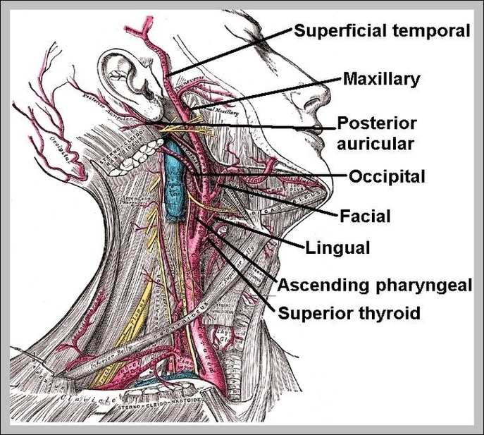
One carotid artery is located on each side of your neck. When your doctor puts their hands on your neck to detect a pulse, they're feeling one of your carotid…
One carotid artery is located on each side of your neck. When your doctor puts their hands on your neck to detect a pulse, they're feeling one of your carotid…
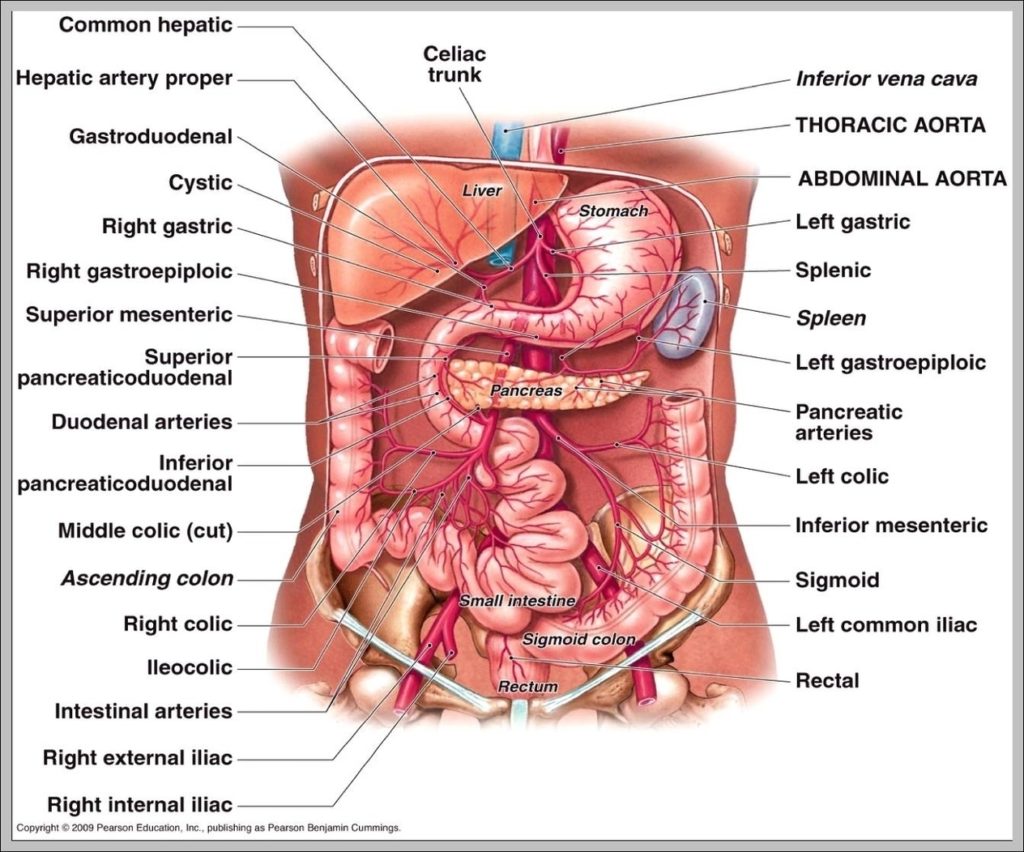
Celiac artery. Dr Sonam Vadera and Dr Donna D'Souza ◉ et al. Celiac artery, also known as the celiac axis or celiac trunk, is a major visceral artery in the…