The arterial supply to the lower limb is a complex network that ensures oxygenated blood reaches the muscles, bones, and skin of the leg and foot. Let’s delve into the intricate anatomy of these arteries, their branches, and their clinical significance.
## 1. Femoral Artery: The Main Artery of the Lower Limb
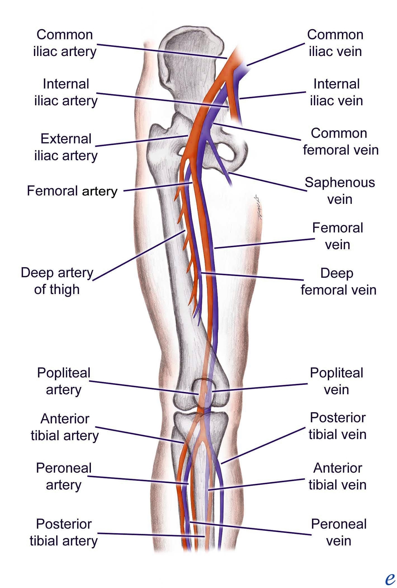
The femoral artery is the primary conduit for blood flow to the lower limb. It originates as a continuation of the external iliac artery, which itself arises from the abdominal aorta. As the external iliac artery crosses under the inguinal ligament, it transforms into the femoral artery and enters the femoral triangle. Here, it gives rise to the profunda femoris artery, which plays a crucial role in supplying the thigh muscles.
### Branches of the Femoral Artery:
1. Perforating Branches: These three or four arteries perforate the adductor magnus muscle, contributing to the blood supply of the medial and posterior thigh.
2. Lateral Femoral Circumflex Artery: Wrapping around the anterior and lateral aspects of the femur, this branch supplies some of the muscles on the outer side of the thigh.
3. Medial Femoral Circumflex Artery: Encircling the posterior side of the femur, it provides blood to the femoral neck and head. In cases of femoral neck fractures, damage to this artery can lead to avascular necrosis of the femoral head.
After exiting the femoral triangle, the femoral artery continues down the anterior thigh within a tunnel called the adductor canal. Along its descent, it supplies the anterior thigh muscles. The journey culminates at the adductor hiatus, an opening in the adductor magnus, through which the femoral artery transitions into the posterior compartment of the thigh, proximal to the knee. At this point, it becomes the popliteal artery.
### Clinical Relevance: Accessing the Femoral Artery
The superficial location of the femoral artery within the femoral triangle makes it easily accessible. Clinicians often use it for various procedures, such as coronary angiography. During this procedure, a catheter is threaded through the femoral artery, allowing visualization of coronary vessels via X-ray after injecting a radio-opaque dye.
## 2. Other Arteries of the Thigh
In addition to the femoral artery, several other vessels contribute to the arterial supply of the lower limb:
### Obturator Artery
– Originates from the internal iliac artery in the pelvic region.
– Descends through the obturator canal to enter the medial thigh.
– Bifurcates into two branches:
– Anterior branch: Supplies the adductor muscles.
– Posterior branch: Provides blood to the hip joint.
### Popliteal Artery
– Derived from the femoral artery.
– Travels behind the knee joint.
– Divides into the anterior tibial artery and the posterior tibial artery.
### Anterior Tibial Artery
– Supplies the anterior compartment of the leg.
– Continues into the foot as the dorsalis pedis artery.
### Posterior Tibial Artery
– Supplies the posterior compartment of the leg.
– Forms the plantar arch in the foot.
## Conclusion
The intricate network of arteries in the lower limb ensures efficient blood supply, nourishing the muscles, bones, and skin. Understanding this vascular system is essential for clinicians and anatomists alike, as it impacts both clinical practice and surgical interventions.
For further exploration, refer to the detailed anatomical diagrams and illustrations available in textbooks and educational resources.