the anterior compartment muscles of the leg. These muscles play a crucial role in movement, stability, and overall function of the lower limb. Without further ado, let’s explore their anatomy, actions, and clinical significance.
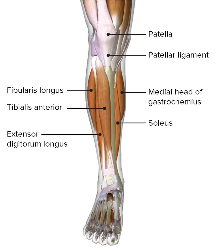
1. Tibialis Anterior:
– Origin: The tibialis anterior muscle originates from the lateral surface of the tibia.
– Attachments: It attaches to the medial cuneiform and the base of metatarsal I.
– Function:
– Dorsiflexion: The tibialis anterior is the strongest dorsiflexor of the foot. It helps lift the foot during walking and prevents it from dragging.
– Inversion: It also contributes to inversion of the foot, which involves turning the sole inward.
– Innervation: The deep fibular nerve supplies the tibialis anterior.
2. Extensor Digitorum Longus:
– Origin: This muscle arises from the lateral condyle of the tibia and the medial surface of the fibula.
– Attachments: Its tendon splits into four and inserts onto the dorsal surface of the lateral four toes.
– Function:
– Toe Extension: The extensor digitorum longus extends the lateral four toes (digits II to V).
– Foot Dorsiflexion: It also assists in dorsiflexion of the foot.
– Innervation: Like the tibialis anterior, it is innervated by the deep fibular nerve.
3. Extensor Hallucis Longus:
– Position: Deep to both the tibialis anterior and extensor digitorum longus.
– Tendon Path: Its tendon emerges from between these two muscles and inserts onto the base of the distal phalanx of the great toe.
– Function:
– Great Toe Extension: The extensor hallucis longus primarily extends the great toe (digit I).
– Foot Dorsiflexion: It also contributes to dorsiflexion of the foot.
– Innervation: Deep fibular nerve.
4. Fibularis Tertius:
– Origin: Arises from the most distal part of the extensor digitorum longus (though it’s not present in all individuals).
– Attachments: Its tendon descends onto the dorsal surface of the fifth metatarsal.
– Function:
– Foot Eversion: The fibularis tertius assists in eversion of the foot, which involves turning the sole outward.
– Foot Dorsiflexion: It also plays a role in dorsiflexion.
– Innervation: Deep fibular nerve.
5. Clinical Relevance: Footdrop:
– Footdrop refers to an inability to dorsiflex the foot at the ankle joint, causing the foot to “drop” under gravity.
– It results from paralysis or weakness of the muscles in the anterior compartment of the leg.
– Typically occurs due to damage to the common fibular nerve, from which the deep fibular nerve arises.
In summary, these anterior compartment muscles work harmoniously to maintain balance, facilitate movement, and prevent mishaps like footdrop. Their intricate interactions make them essential players in our daily activities, whether we’re walking, running, or simply standing. Next time you take a step, remember the unsung heroesthe anterior leg muscles! ????????.
our