
Deltoid muscle (Musculus deltoideus) The deltoid is a thick, triangular shoulder muscle. It gets its name because of its similar shape to the Greek letter ‘delta’ (Δ). The muscle has a wide origin spanning the clavicle, acromion and spine of scapula.
Origins and Insertion. The very broad origin of the deltoid actually means that functionally, the muscle can be broken down into three separate parts: Unlike other muscles, for example the triceps brachii (tri = three, ceps = heads), the name of the deltoid muscle does not indicate this possible functional division.
Muscle Testing. To properly test the function of the deltoid and the axillary nerve, the arm must be beyond 15 degrees of abduction. Once the arm is in this position, the patient then pushes against resistance. If the muscle is functioning properly, contraction of the muscle should be felt near the acromion of the shoulder.
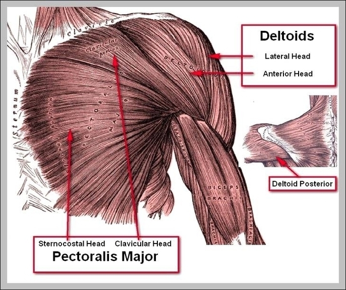
Deltoid Diagram Image
Posted inDiagrams
Deltoid Diagram Image
Post navigation
Previous Post
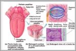 Location Of Taste Buds Image
Next Post
Human Organ Picture 2 Image 