
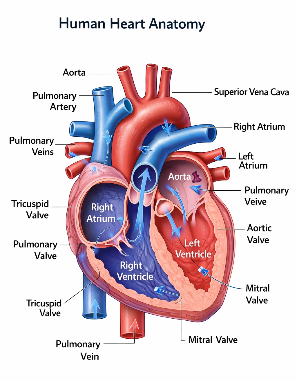
The human heart anatomy diagram shows how this powerful muscle works as the body’s central pump, constantly circulating blood to keep us alive. The heart is divided into four main chambers: the right atrium and right ventricle on one side, and the left atrium and left ventricle on the other. Blood low in oxygen enters the right atrium, moves into the right ventricle, and is then sent to the lungs through the pulmonary artery to pick up oxygen. Once oxygen-rich, the blood returns to the left atrium, flows into the left ventricle, and is pumped out through the aorta to supply the entire body. This continuous flow is what keeps organs functioning and energy levels stable.
Another key part of human heart anatomy is the system of valves that ensure blood moves in the correct direction. The tricuspid, pulmonary, mitral, and aortic valves open and close with each heartbeat, preventing any backward flow. The walls of the heart, especially the left ventricle, are thick and strong because they need to pump blood with enough force to reach all parts of the body. When you look at a labeled heart diagram, it becomes much easier to understand how all these parts work together in a smooth, coordinated way. This is why heart anatomy diagrams are so popular for students, educators, and anyone wanting a clear, visual explanation of how the cardiovascular system works.
