
The small intestine’s histology maximizes surface area for digestion and absorption across its three segmentsduodenum, jejunum, ileumwith tall villi covered in microvilli on enterocytes forming the brush border loaded with enzymes like lactase and peptidases. Goblet cells increase distally for mucus, Paneth cells in crypt bases secrete defensins against bacteria, and Brunner’s glands in duodenum produce alkaline mucus to neutralize acid. Peyer’s patches in ileum monitor antigens for immunity. Circular folds called plicae circulares slow flow, submucosa supports, muscularis externa propels via peristalsis, and serosa or adventitia finishes it. Nutrient uptake occurs via transporters, with lipids packaged into chylomicrons entering lacteals.
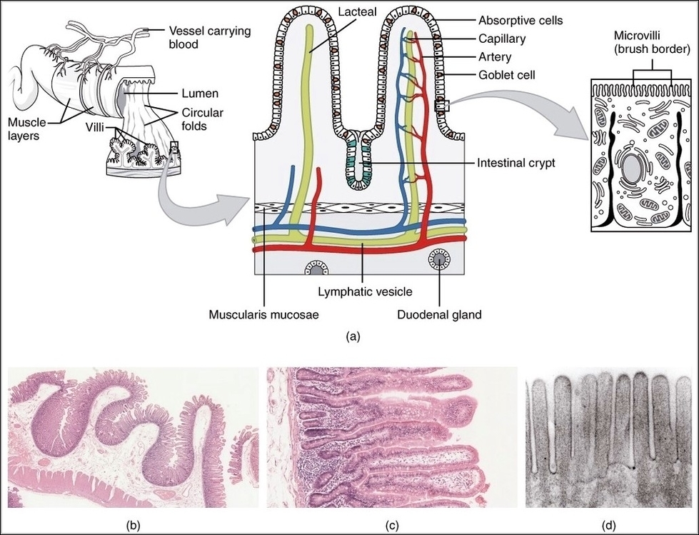
Histology Small IntestinesN
Posted inOrgans
Histology Small IntestinesN
Post navigation
Previous Post
 Elbow 3D Diagram