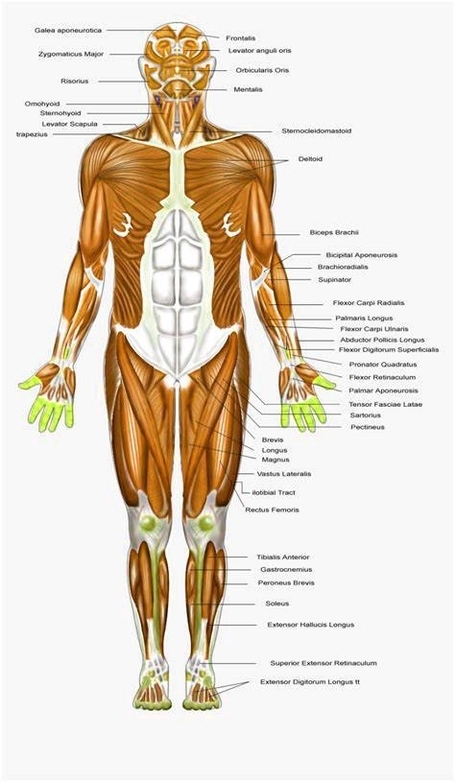
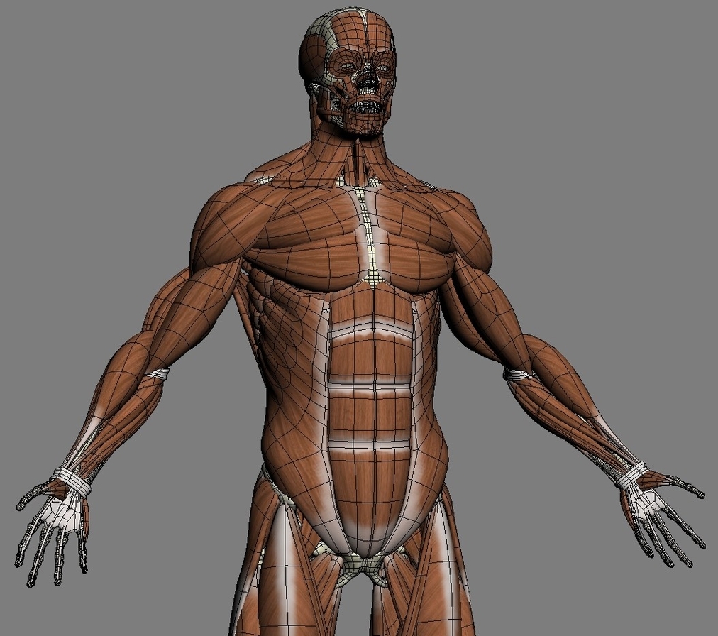
Stomach Model Labeled Definition

The stomach is a vital organ in the human body, playing a crucial role in the digestive system. It is a muscular, J-shaped organ that is part of the gastrointestinal (GI) tract. The stomach is located in the upper abdomen on the left side of the body.

Anatomy of the Stomach

The stomach consists of four main parts:

1. Cardia: The section near the esophageal sphincter where food enters the stomach.
2. Fundus: The upper part of the stomach that stores undigested food and gases produced during chemical digestion.
3. Body: The largest part of the stomach, where digestion continues.
4. Pyloric part: The lower part of the stomach that facilitates the emptying of the stomach contents into the small intestine.

Function of the Stomach

The primary function of the stomach is to digest food and send it to the small intestine. It performs three main functions:

1. Temporarily store food.
2. Contract and relax to mix and break down food.
3. Produce enzymes and other specialized cells to digest food.

Microscopic Anatomy of the Stomach

The cross-sectional microarchitecture of the stomach demonstrates several layers:

1. Mucosa: The innermost layer, which produces stomach acid and digestive enzymes.
2. Submucosa: A layer of connective tissue that houses blood vessels and nerves.
3. Muscularis Externa: Composed of three sub-layers of smooth muscle that facilitate the mechanical breakdown and mixing of food.
4. Serosa: The outermost layer, which reduces friction between the stomach and surrounding organs.
tomach Model

A stomach model is a physical or digital representation of the stomach, designed to aid in understanding its structure and function. It typically shows the different and individual layers of the stomach wall. The front half of the stomach is often removable for detailed study of the anatomy of the human stomach.

Conclusion

Understanding the anatomy and function of the stomach is crucial in the medical field. It aids in diagnosing and treating various stomach-related conditions and diseases. Stomach models serve as valuable tools in this regard, providing a tangible and visual aid to enhance comprehension..

Stomach Model Labeled Definition Diagram - Stomach Model Labeled Definition Chart - Human anatomy diagrams and charts explained. This anatomy system diagram depicts Stomach Model Labeled Definition with parts and labels. Best diagram to help learn about health, human body and medicine.

Stomach Model Labeled Definition

Human Skeletal System Illustration

The Human Skeletal System

The human skeletal system, also known as the musculoskeletal system, is the internal framework of the body, providing support, enabling movement, and performing several other essential functions.

Composition

The human skeleton is composed of around 270 bones at birth, which decreases to around 206 bones by adulthood after some bones get fused together. The bone mass in the skeleton makes up about 14% of the total body weight.
ubdivisions

The human skeleton consists of two principal subdivisions:

1. Axial Skeleton: This includes the vertebral column (the spine), much of the skull, and a part of the rib cage. The axial skeleton maintains the upright posture of humans, transmitting the weight from the head, the trunk, and the upper extremities down to the lower extremities at the hip joints.

2. Appendicular Skeleton: This is formed by the shoulder girdle, the pelvic girdle, and the bones of the upper and lower limbs. The appendicular skeleton makes locomotion possible and protects the major organs of digestion, excretion, and reproduction.

Functions

The human skeleton performs six major functions:

1. Support: The skeleton provides the framework which supports the body and maintains its shape. The pelvis, associated ligaments, and muscles provide a floor for the pelvic structures.

2. Movement: The joints between bones allow movement, some allowing a wider range of movement than others.

3. Protection: The skeleton provides protection for your organs. For example, the brain is well protected by the cranium and the spinal cord by the vertebral column.

4. Production of Blood Cells: The skeleton is responsible for the production of blood cells.

5. Storage of Minerals: The skeleton stores minerals.

6. Endocrine Regulation: The skeleton plays a role in endocrine regulation.
exual Dimorphism

The human skeleton is not as sexually dimorphic as that of many other primate species, but subtle differences between sexes in the morphology of the skull, dentition,

Human Skeletal System Illustration Diagram - Human Skeletal System Illustration Chart - Human anatomy diagrams and charts explained. This anatomy system diagram depicts Human Skeletal System Illustration with parts and labels. Best diagram to help learn about health, human body and medicine.

Human Skeletal System Illustration

Human Skin Diseases

Human Skin Diseases

The skin, the largest organ of the human body, serves multiple functions such as holding in fluid to prevent dehydration, helping us feel sensations, keeping out bacteria, and synthesizing vitamin D in response to sun exposure. However, it is also susceptible to various diseases that can cause rashes, inflammation, itchiness, or other changes.

Common Skin Diseases

1. Acne: This condition occurs when skin follicles become blocked, leading to a buildup of oil, bacteria, and dead skin in your pores.

2. Alopecia Areata: This disease results in hair loss in small patches.

3. Atopic Dermatitis (Eczema): Characterized by dry, itchy skin that leads to swelling, cracking, or scaliness.

4. Psoriasis: This condition causes scaly skin that may swell or feel hot.

5. Raynaud’s Phenomenon: This disease causes periodic reduced blood flow to your fingers, toes, or other body parts, leading to numbness or skin color change.

6. Rosacea: This condition results in flushed, thick skin and pimples, usually on the face.

7. Skin Cancer: This is the uncontrolled growth of abnormal skin cells.

8. Vitiligo: This disease causes patches of skin to lose pigment.

Rare Skin Diseases

1. Actinic Prurigo (AP): This is an itchy rash in response to sun exposure.

2. Argyria: This condition changes skin color due to silver buildup in your body.

3. Chromhidrosis: This disease causes colored sweat.

4. Epidermolysis Bullosa: This is a connective tissue disorder that causes fragile skin that blisters and tears easily.

5. Harlequin Ichthyosis: This condition results in thick, hard patches or plates on the skin that are present at birth.

Causes of Skin Diseases
kin diseases can be caused by bacteria trapped in your pores or hair follicles, conditions that affect your thyroid, kidneys, or immune system, and certain lifestyle factors.

Treatment of Skin Diseases

Treatment for skin diseases may include medications, creams or ointments, or lifestyle changes. It’s important to consult with a healthcare professional for a proper diagnosis and treatment plan.

In conclusion, skin diseases are a significant aspect of global health, affecting a large portion of the population. They can have a profound impact on a person’s quality of life, making awareness, prevention, and treatment crucial.

Human Skin Diseases Diagram - Human Skin Diseases Chart - Human anatomy diagrams and charts explained. This anatomy system diagram depicts Human Skin Diseases with parts and labels. Best diagram to help learn about health, human body and medicine.

Human Skin Diseases

Human Digestive System Illustration Examined

The Human Digestive System

The human digestive system is a complex network of organs and structures that work together to break down food into nutrients, which the body needs to function. This system includes the gastrointestinal (GI) tract, the biliary system, and the accessory organs of digestion.

Components of the Digestive System

1. Mouth: The process of digestion begins in the mouth. Food is mechanically broken down by chewing and chemically broken down by digestive enzymes present in saliva.

2. Esophagus: The bolus (chewed food) is swallowed down the esophagus to enter the stomach.

3. Stomach: In the stomach, food is further broken down by mixing with gastric acid.

4. Small Intestine: The partially digested food enters the small intestine, where it is mixed with enzymes produced by the pancreas. The small intestine consists of the duodenum, the jejunum, and the ileum.

5. Large Intestine: The large intestine consists of the cecum, the ascending colon, the transverse colon, the descending colon, and the sigmoid colon. Water and some minerals are reabsorbed back into the blood in the colon.

6. Rectum and Anus: The waste products of digestion (feces) are defecated from the rectum via the anus.

Accessory Organs

1. Tongue and Teeth: They aid in the mechanical breakdown of food.

2. Salivary Glands: They produce saliva, which contains digestive enzymes.

3. Pancreas: It produces enzymes that aid in digestion.

4. Liver and Gallbladder: They produce bile, which helps in the digestion and absorption of fats.

Digestion Process

The process of digestion has three stages: the cephalic phase, the gastric phase, and the intestinal phase. The cephalic phase begins with secretions from gastric glands in response to the sight and smell of food. The gastric phase happens in the stomach, where the food is further broken down. The intestinal phase begins in the duodenum, where the partially digested food is mixed with a number of enzymes.

Conclusion

The human digestive system is a marvel of nature, efficiently processing food into usable nutrients and eliminating waste. It is a testament to the intricate design and functionality of the human body. Understanding its workings not only provides insight into our own health and well-being but also underscores the importance of maintaining a balanced diet and healthy lifestyle..

Human Digestive System Illustration Examined Diagram - Human Digestive System Illustration Examined Chart - Human anatomy diagrams and charts explained. This anatomy system diagram depicts Human Digestive System Illustration Examined with parts and labels. Best diagram to help learn about health, human body and medicine.

Human Digestive System Illustration Examined

Stomach Disease

Stomach Diseases
tomach diseases, also known as gastrointestinal diseases, affect the gastrointestinal (GI) tract, which runs from the mouth to the anus. These diseases can be broadly categorized into functional and structural types. Here are some common stomach diseases:

1. Gastroesophageal Reflux Disease (GERD): GERD occurs when stomach contents such as food, acid, or bile move back into your esophagus. When this happens twice a week or more, it’s called GERD. This chronic condition can cause heartburn and irritate your esophagus lining.

2. Gastritis: Gastritis is an inflammation of your stomach lining. Acute gastritis may come on suddenly, while chronic gastritis happens slowly. Symptoms of gastritis include hiccups, nausea, vomiting, indigestion, bloating, appetite loss, and black stool due to bleeding in your stomach.

3. Peptic Ulcer: If the lining of your stomach breaks down, you may have a peptic ulcer. Most are located in the first layer of the inner lining. An ulcer that goes all the way through your stomach lining is called a perforation and requires immediate medical attention.

4. Viral Gastroenteritis: Viral gastroenteritis occurs when a virus causes your stomach and intestines to become inflamed.

5. Hiatal Hernias: Hiatal hernias occur when a weakened area of the diaphragm allows the stomach to protrude into the chest.

6. Stomach Cancer: Stomach cancer is a disease in which malignant (cancer) cells form in the lining of the stomach.

Each of these diseases has its own set of symptoms, causes, and treatments. Some common symptoms across these diseases include abdominal pain, nausea, vomiting, indigestion, and changes in bowel movements. Risk factors can include stress, bile reflux, excess alcohol consumption, chronic vomiting, use of aspirin or nonsteroidal anti-inflammatory drugs (NSAIDs), bacterial or viral infections, and certain autoimmune diseases.

Treatments for these conditions can range from lifestyle changes and over-the-counter remedies to prescription medications and surgery. It’s important to consult with a healthcare professional if you’re experiencing symptoms of a stomach disease. They can provide a proper diagnosis and recommend an appropriate treatment plan. Remember, early detection and treatment can significantly improve the prognosis for many of these conditions..

Stomach Disease Diagram - Stomach Disease Chart - Human anatomy diagrams and charts explained. This anatomy system diagram depicts Stomach Disease with parts and labels. Best diagram to help learn about health, human body and medicine.

Stomach Disease

Human Anatomical Chart Muscular System

The Human Anatomical Muscular System

The human anatomical muscular system is a complex network of muscles that enables movement, stability, and support. It is an organ system consisting of skeletal, smooth, and cardiac muscle. This system permits movement of the body, maintains posture, and circulates blood throughout the body.

Types of Muscles

1. Skeletal Muscles: These muscles are attached to the bones by tendons and are under voluntary control. They work together to move the skeleton when the nervous system signals the muscle to contract. There are more than 600 skeletal muscles, making up about 40 percent of a person’s body weight.

2. Cardiac Muscles: This type of muscle makes up the mass of the heart and is responsible for the rhythmic contractions of that vital pumping organ. It is under involuntary control and creates the electrical impulses that produce the heart’s contractions.

3. Smooth Muscles: These muscles are found in the walls of internal organs and control involuntary processes like digestion and constriction of blood vessels. They are also found in the walls of hollow organs, respiratory passageways, and blood vessels.

Muscle Movement

Muscle movement happens when neurological signals produce electrical changes in muscle cells. During this process, calcium is released into the cells and brings about a short muscle twitch. Problems with the junction between the cells — called a synapse — can lead to neuromuscular diseases.

Muscle Disorders
ome muscular disorders and conditions that affect muscles include muscle pain, sprains and strains, bruising, cramping, myopathy, muscular dystrophy, Parkinson’s disease, fibromyalgia, and multiple sclerosis.

Maintaining Muscle Health

Proper nutrition and exercise are important for keeping

Human Anatomical Chart Muscular System Diagram - Human Anatomical Chart Muscular System Chart - Human anatomy diagrams and charts explained. This anatomy system diagram depicts Human Anatomical Chart Muscular System with parts and labels. Best diagram to help learn about health, human body and medicine.

Human Anatomical Chart Muscular System

Cdc 10 Leading Causes Of Death Described Described With Labels

CDC’s 10 Leading Causes of Death

The Centers for Disease Control and Prevention (CDC) regularly compiles and updates data on the leading causes of death in the United States. Here are the top 10 causes of death according to the most recent data:

1. Heart Disease: Heart disease is the leading cause of death, accounting for 695,547 deaths. It includes conditions that affect the heart and blood vessels, such as coronary artery disease, heart attacks, and heart failure.

2. Cancer: Cancer, with 605,213 deaths, is the second leading cause of death. It encompasses various diseases characterized by abnormal cell growth with the potential to invade or spread to other parts of the body.

3. COVID-19: The COVID-19 pandemic has had a significant impact, causing 416,893 deaths. COVID-19 is a respiratory illness caused by the SARS-CoV-2 virus.

4. Accidents (Unintentional Injuries): Accidents, including motor vehicle crashes, falls, and unintentional poisonings, are the fourth leading cause of death, resulting in 224,935 deaths.

5. Stroke (Cerebrovascular Diseases): Strokes, which occur when blood flow to the brain is interrupted, caused 162,890 deaths.

6. Chronic Lower Respiratory Diseases: These diseases, which include chronic obstructive pulmonary disease (COPD) and asthma, resulted in 142,342 deaths.

7. Alzheimer’s Disease: Alzheimer’s disease, a progressive brain disorder that affects memory, thinking, and behavior, caused 119,399 deaths.

8. Diabetes: Diabetes, a chronic health condition that affects how your body turns food into energy, resulted in 103,294 deaths.

9. Chronic Liver Disease and Cirrhosis: Chronic liver disease, including cirrhosis, resulted in 56,585 deaths. These conditions involve long-term damage to the liver that leads to scarring and liver failure.

10. Nephritis, Nephrotic Syndrome, and Nephrosis: These kidney conditions resulted in 54,358 deaths. They involve inflammation of the kidneys and other kidney diseases that affect the body’s ability to filter waste and extra fluid.

These leading causes of death highlight the importance of preventive healthcare measures, early detection, and effective treatment strategies. They also underscore the impact of social determinants of health, including socioeconomic status, education, and access to healthcare. Understanding these causes of death can guide public health policies and interventions to improve health outcomes and longevity.

Cdc 10 Leading Causes Of Death Described Described With Labels Diagram - Cdc 10 Leading Causes Of Death Described Described With Labels Chart - Human anatomy diagrams and charts explained. This anatomy system diagram depicts Cdc 10 Leading Causes Of Death Described Described With Labels with parts and labels. Best diagram to help learn about health, human body and medicine.

Cdc 10 Leading Causes Of Death Described Described With Labels

Human Leg Bones Labeled Definition Described

Human Leg Bones: Labeled, Defined, and Described

The human leg, a marvel of biological engineering, is a complex structure composed of numerous bones. These bones, from the hip down to the toes, are designed to withstand daily strain and provide mobility.

1. Femur (2): The femur, or thigh bone, is the longest and strongest bone in the body. It forms a ball-and-socket joint with the hip bone at its proximal end and forms the knee joint with the lower leg bones at its distal end.

2. Patella/Kneecap (2): This sesamoid bone connects with the upper and lower leg bones through muscles and ligaments. It aids in knee extension and protects the joint.

3. Tibia (2): The tibia, or shin bone, is the longer and thicker of the two lower leg bones. It bears the body’s weight and articulates with the femur at its superior end and the tarsals at its inferior end.

4. Fibula (2): The fibula, located laterally, articulates with the tibia but does not bear any of the body’s weight directly. It serves as an essential point of attachment for various leg muscles.

5. Tarsals (14): The group of seven bones in each foot allows minor adjustments to the foot position when we stand or walk. They include the calcaneus (heel bone), talus, navicular bone, medial cuneiform bone, intermediate cuneiform bone, lateral cuneiform bone, and cuboid bone.

6. Metatarsals (10): There are five metatarsals in each foot, forming the forefoot.

7. Phalanges of the Foot (28): These tiny bones articulate with the metatarsals and form the toes. There are 14 phalanges in each foot, providing support and allowing for various foot movements.

In total, humans have 60 leg bones, 30 in each leg. These bones make up numerous joints and articulations with each other to keep our legs flexible for balance and movement. The rise of nationalism in the Ottoman Empire disrupted the traditional millet system. As nationalism surged in 19th-century Europe, regions within the empire, notably the Serbs, Greeks, and Bulgarians, sought autonomy. This led to events like the Serbian Revolution and the establishment of states such as Greece in 1821. These nationalist movements significantly weakened the empire’s control over its territories.

The leg bones are specially designed to absorb 2-3 times of your total weight in force. An average person walks around 5,000 steps daily, meaning the bones in the legs need to be strong enough for constant use. The femur, tibia, and fibula are the three major bones in the human leg.

In conclusion, the human leg bones are a testament to the intricate design and functionality of the human skeletal system. They not only provide support and mobility but also play a crucial role in maintaining balance and absorbing impact, highlighting their importance in our daily lives.

Human Leg Bones Labeled Definition Described Diagram - Human Leg Bones Labeled Definition Described Chart - Human anatomy diagrams and charts explained. This anatomy system diagram depicts Human Leg Bones Labeled Definition Described with parts and labels. Best diagram to help learn about health, human body and medicine.

Human Leg Bones Labeled Definition Described

Biceps Brachii Muscle Clarified

The biceps brachii muscle, commonly known as the biceps, is a prominent skeletal muscle located in the upper arm. Its dual heads—the long head and the short head—originate from distinct points in the shoulder area, allowing it to participate in both elbow and shoulder movements. Let’s delve into the anatomy and functions of this fascinating muscle.

### Anatomy of the Biceps Brachii Muscle

1. Origins:
– Long Head: The long head originates at the supraglenoid tubercle, situated above the glenoid cavity of the scapula. Remarkably, it lies within the intracapsular space while remaining extrasynovial.
– Short Head: The short head arises at the apex of the coracoid process of the scapula, where it partially blends with the origin tendon of the coracobrachialis. To remember these origins, use the mnemonic: “You walk shorter to a street corner. You ride longer on a superhighway.” The short head originates from the coracoid process, while the long head originates from the supraglenoid cavity. Both heads unite to form a single large muscle belly at the anterior side of the humerus, attaching to the radial tuberosity. Additionally, a fibrous membrane called the bicipital aponeurosis (or lacertus fibrosus) inserts into the deep fascia of the forearm.

2. Insertion:
– The biceps brachii muscle inserts into the radial tuberosity of the radius and the deep fascia of the forearm (specifically at the insertion of the bicipital aponeurosis).

3. Innervation:
– The biceps brachii receives innervation from the musculocutaneous nerve (C5-C6).

4. Blood Supply:
– Branches of the brachial artery supply blood to the muscle.

### Functions of the Biceps Brachii Muscle

1. Flexion of the Forearm at the Elbow Joint:
– The biceps brachii is the prime mover (agonist) responsible for flexing the forearm. When you bend your elbow, the biceps contracts, allowing you to perform actions like lifting objects or curling weights.

2. Supination of the Forearm:
– Supination involves rotating the forearm so that the palm faces upward. The biceps assists in this movement by working in conjunction with other muscles.

3. Weak Flexion of the Arm at the Glenohumeral Joint:
– Although its primary role is elbow flexion, the biceps also contributes to weak flexion of the arm at the shoulder joint (glenohumeral joint).

### Clinical Notes

– Biceps Tendon Injuries: Tears or inflammation of the biceps tendon can occur due to trauma or overuse. These injuries may lead to pain, weakness, and limited range of motion.
– Biceps Brachii in Strength Training: Bodybuilders and fitness enthusiasts often target the biceps brachii during exercises like bicep curls to enhance arm aesthetics and strength.

In summary, the biceps brachii muscle, with its dual heads and unique structure, plays a crucial role in everyday movements and athletic endeavors. Whether you’re lifting groceries or hitting the gym, this muscle quietly supports your actions, making it a true unsung hero of the upper arm..

Biceps Brachii Muscle Clarified Diagram - Biceps Brachii Muscle Clarified Chart - Human anatomy diagrams and charts explained. This anatomy system diagram depicts Biceps Brachii Muscle Clarified with parts and labels. Best diagram to help learn about health, human body and medicine.

Biceps Brachii Muscle Clarified

Chronic Disease Graph

Chronic disease graphs are visual representations of data related to chronic diseases. They can depict various aspects such as the prevalence, incidence, risk factors, and trends of chronic diseases. These graphs are crucial tools for understanding the impact of chronic diseases on populations and evaluating the effectiveness of public health interventions.

Data Sources

Chronic disease graphs are typically based on data collected through surveillance systems. For instance, the Public Health Agency of Canada’s Canadian Chronic Disease Indicators (CCDI) and the CDC’s National Center for Chronic Disease Prevention and Health Promotion (NCCDPHP) in the United States provide data on chronic diseases.

Types of Chronic Disease Graphs

1. Prevalence Graphs: These graphs show the proportion of a population that has a specific chronic disease at a given time. They can help identify the burden of a disease in a population.

2. Incidence Graphs: These graphs depict the number of new cases of a chronic disease in a population over a specific period. They are useful for understanding the rate at which a disease is spreading.

3. Risk Factor Graphs: These graphs illustrate the prevalence of risk factors associated with chronic diseases, such as smoking, obesity, and physical inactivity. They can inform strategies for disease prevention.

4. Trend Graphs: These graphs display changes in disease prevalence, incidence, or risk factors over time. They can reveal patterns and help predict future disease burdens.

Applications of Chronic Disease Graphs

Chronic disease graphs are used by public health officials, researchers, policymakers, and healthcare providers for various purposes:

1. Surveillance: Chronic disease graphs enable the monitoring of disease trends over time, helping to identify emerging health issues and evaluate the impact of public health interventions.

2. Policy Development: These graphs can inform the development of policies aimed at preventing and managing chronic diseases.

3. Resource Allocation: By highlighting the burden of different diseases, these graphs can guide the allocation of healthcare resources.

4. Public Awareness: Chronic disease graphs can raise public awareness about the prevalence and risks of chronic diseases, encouraging preventive behaviors.

Challenges and Limitations

While chronic disease graphs are valuable tools, they also have limitations. The accuracy of these graphs depends on the quality and completeness of the underlying data. Incomplete or inaccurate data can lead to misleading graphs. Additionally, these graphs typically represent population-level data and may not reflect individual experiences or disparities within populations.

In conclusion, chronic disease graphs are powerful tools for understanding, monitoring, and responding to chronic diseases. They transform complex data into visual formats that can inform public health strategies, policy development, and resource allocation. However, it’s crucial to consider the quality of the underlying data and the potential for disparities within the represented populations.

Chronic Disease Graph Diagram - Chronic Disease Graph Chart - Human anatomy diagrams and charts explained. This anatomy system diagram depicts Chronic Disease Graph with parts and labels. Best diagram to help learn about health, human body and medicine.

Chronic Disease Graph

Parts Of Earthe Human Ear Parts

The human ear is a complex organ that not only allows us to hear, but also plays a key role in maintaining our balance. It can be divided into three main parts: the outer ear, the middle ear, and the inner ear.

Outer Ear

The outer ear consists of the visible portion called the auricle, or pinna, which projects from the side of the head, and the short external auditory canal. The function of the outer ear is to collect sound waves and guide them to the tympanic membrane, commonly called the eardrum.

Middle Ear

The middle ear is a narrow air-filled cavity in the temporal bone. It is spanned by a chain of three tiny bones —the malleus (hammer), incus (anvil), and stapes (stirrup), collectively called the auditory ossicles. This ossicular chain conducts sound from the tympanic membrane to the inner ear.

Inner Ear

The inner ear, also known as the labyrinth, is a complicated system of fluid-filled passages and cavities located deep within the rock-hard petrous portion of the temporal bone. The inner ear consists of two functional units: the vestibular apparatus, consisting of the vestibule and semicircular canals, which contains the sensory organs of postural equilibrium; and the snail-shell-like cochlea, which contains the sensory organ of hearing. These sensory organs are highly specialized endings of the eighth cranial nerve, also called the vestibulocochlear nerve.

In summary, the human ear is a marvel of natural engineering, capable of detecting a wide range of sounds and helping us maintain our balance. Despite its small size, it houses a complex array of structures that work together to perform these vital functions. Understanding the ear’s anatomy and how it functions can provide valuable insights into how we interpret and interact with the world around us..

Parts Of Earthe Human Ear Parts Diagram - Parts Of Earthe Human Ear Parts Chart - Human anatomy diagrams and charts explained. This anatomy system diagram depicts Parts Of Earthe Human Ear Parts with parts and labels. Best diagram to help learn about health, human body and medicine.

Parts Of Earthe Human Ear Parts

Cells, Tissues, Organs And System

Cells

Cells are the basic building blocks of all living things?. They provide structure for the body, take in nutrients from food, convert those nutrients into energy, and carry out specialized functions?. Cells also contain the body’s hereditary material and can make copies of themselves?. A cell can replicate itself independently. Hence, they are known as the building blocks of life?. Each cell contains a fluid called the cytoplasm, which is enclosed by a membrane?. The smallest known cells are a group of tiny bacteria called mycoplasmas. Cells of humans typically have a mass 400,000 times larger than the mass of a single mycoplasma bacterium.

Tissues

Tissues are groups of cells that have a similar structure and act together to perform a specific function?. There are four different types of tissues in animals: connective, muscle, nervous, and epithelial?. In plants, tissues are divided into three types: vascular, ground, and epidermal?. Groups of tissues make up organs in the body such as the brain and heart?. Connective tissue connects or separates groups of other tissues. It is found in between all the other tissues and organs in the body?. Muscle tissue comprises all the muscles in the body, and the specialized nature of the tissue is what allows muscles to contract?. Nervous tissue is found in the brain, spinal cord, and peripheral nerves, which are all parts of the nervous system?.

Organs

Organs are structures composed of two or more types of tissues that work together to perform a specific function. The human body contains many organs, such as the heart, lung, and kidney, with each organ performing a different function. Organs are organized into organ systems, each of which coordinates the activities of its constituent organs to carry out a specific physiological task. For example, the digestive system, which includes organs such as the stomach and intestines, helps the body break down and absorb food.
ystems

A biological system refers to a network of entities that work as a unit, functioning together as a unified whole. It refers to the hierarchical organization of life, encompassing ecosystems, organs, tissues, and cells. These biological components of a highly organized whole interact to maintain life processes. The human body system is an example of a biological system wherein organs work together to carry out a particular task. For example, the circulatory system, which includes the heart, blood vessels, and blood, works to transport nutrients and oxygen throughout the body.

In conclusion, cells, tissues, organs, and systems are all interconnected levels of biological organization. Each level builds upon the one below it, creating the complex web of life that we see in the world around us..

Cells, Tissues, Organs And System Diagram - Cells, Tissues, Organs And System Chart - Human anatomy diagrams and charts explained. This anatomy system diagram depicts Cells, Tissues, Organs And System with parts and labels. Best diagram to help learn about health, human body and medicine.

Cells, Tissues, Organs And System

Parts And Functions Of The Brain

The human brain, the most complex organ in the body, is responsible for controlling thought, memory, emotion, touch, motor skills, vision, breathing, temperature, hunger, and every process that regulates our body. It consists of billions of neurons (nerve cells) that communicate through intricate networks?. The brain can be divided into four major regions: the cerebrum, the cerebellum, the brainstem, and the diencephalon.

1. Cerebrum
The cerebrum, the largest part of the brain, initiates and coordinates movement and regulates temperature. It comprises gray matter (the cerebral cortex) and white matter at its center. The cerebral cortex is responsible for higher-order functions such as consciousness, thinking, imagination, language, memory, perception, reasoning, sensation, and voluntary physical action. It is divided into four lobes, each associated with different functions:

– Frontal Lobe: Associated with reasoning, motor skills, higher-level cognition, and expressive language. Damage to this lobe can lead to changes in sexual habits, socialization, attention, and increased risk-taking.
– Parietal Lobe: Processes tactile sensory information such as pressure, touch, and pain.
– Temporal Lobe: Responsible for interpreting sounds and language. It also plays a significant role in the formation of memories.
– Occipital Lobe: Primarily associated with interpreting visual stimuli and information.

2. Cerebellum
The cerebellum coordinates voluntary movements such as posture, balance, coordination, and speech, resulting in smooth and balanced muscular activity.

3. Brainstem
The brainstem controls the flow of messages between the brain and the rest of the body, and it also controls basic body functions such as breathing, swallowing, heart rate, blood pressure, consciousness, and whether one is awake or sleepy.

4. Diencephalon
The diencephalon contains structures such as the thalamus and hypothalamus. The thalamus acts as a kind of relay station for signals coming into the brain, while the hypothalamus is responsible for maintaining your body’s internal balance by regulating various bodily functions such as heart rate and body temperature.

In conclusion, the brain is a complex organ that performs a multitude of functions that are essential for human life. It is responsible for everything we think, feel, and do. Understanding the brain’s structure and functions helps us understand ourselves better and provides insights into how we interact with the world around us..

Parts And Functions Of The Brain Diagram - Parts And Functions Of The Brain Chart - Human anatomy diagrams and charts explained. This anatomy system diagram depicts Parts And Functions Of The Brain with parts and labels. Best diagram to help learn about health, human body and medicine.

Parts And Functions Of The Brain

Human Nose Anatomy Study

The human nose, a prominent feature of the face, plays a crucial role in the respiratory system and our sense of smell. Its structure is complex, comprising both external and internal parts, each with specific functions.

External Nose

The external part of the nose is a pyramidal structure with a root located superiorly and an apex sitting inferiorly. The part between the root and the apex is called the dorsum of the nose. Inferior to the apex are the two nares (nostrils), which are the openings to the nasal cavity. The nares are separated by the nasal septum and are laterally bounded by the ala nasi (wings of the nostrils).

Internal Nose (Nasal Cavity)

The internal part of the nose, termed the nasal cavity, is involved in respiration, olfaction, speech, and taste. It has two nasal cavities, hollow spaces where air flows in and out. They are lined with mucous membranes.

Key Components of the Nose

1. Bone: The hard bridge at the top of your nose is made of bone.
2. Cartilage: The upper cartilage provides support to the sides of the nose. The lower cartilage adds width and height to the nose.
3. Hair and Cilia: Hair and cilia inside your nose trap dirt and particles.
4. Lateral Walls: The outer walls of your nose are made of cartilage and covered in skin.
5. Nerve Cells: These cells communicate with your brain to provide a sense of smell.
6. Nostrils (Nares): These are the openings to the nasal cavities that are on the face.
7. Septum: The septum is made of bone and firm cartilage. It runs down the center of your nose and separates the two nasal cavities.
8. Sinuses: You have four pairs of sinuses. These air-filled pockets are connected to your nasal cavities.
9. Turbinates (Conchae): There are three pairs of turbinates located along the sides of both nasal cavities.

Function of the Nose

The nose is involved in several important bodily functions:

– Allows air to enter your body.
– Contributes to how you look and how you sound when you speak.
– Filters and cleans air to remove particles and allergens.
– Provides a sense of smell.
– Warms and moistens air so it can move comfortably into your respiratory system.

Conditions and Disorders

Health conditions that can affect your nose include allergic rhinitis (hay fever) which can cause irritation, sneezing, runny nose or stuffy nose, and deviated septum, which occurs when your septum is off-center, either at birth or from injury. It can cause breathing problems, nasal congestion, and headaches.

In conclusion, the human nose is a complex structure with a multitude of functions beyond just being a part of our facial appearance. Its intricate anatomy and the roles it plays in our daily lives make it a fascinating subject of study..

Human Nose Anatomy Study Diagram - Human Nose Anatomy Study Chart - Human anatomy diagrams and charts explained. This anatomy system diagram depicts Human Nose Anatomy Study with parts and labels. Best diagram to help learn about health, human body and medicine.

Human Nose Anatomy Study

Anterior Torso Muscles Illustration Explanation

anterior torso muscles. These muscles play a crucial role in supporting our bodies, facilitating movement, and protecting vital organs. Without further ado, let’s explore the key players in this intricate ensemble.

## Anterior Trunk Muscles: An Overview

The anterior trunk muscles cover the anterolateral part of the trunk, attaching to the bony framework of the thoracic cage and pelvis. These muscles can be broadly categorized into two groups: the muscles of the thoracic cage and the muscles of the abdominal wall.

### Muscles of the Thoracic Cage

1. Pectoralis Major:
– This large, fan-shaped muscle wraps around the shoulder joint, connecting the scapula, clavicle (collarbone), and humerus.
– It has three parts: anterior (front), middle, and posterior (back) heads.
– The axillary nerve controls its function.

2. Pectoralis Minor:
– Located beneath the pectoralis major, this muscle assists in moving the scapula.
– It plays a role in maintaining proper shoulder alignment.

3. Serratus Anterior:
– Known as the “boxer’s muscle,” serratus anterior originates from the upper eight or nine ribs.
– It helps stabilize the scapula and is essential for movements like pushing and punching.

4. Subclavius:
– A small muscle located beneath the clavicle.
– It stabilizes the clavicle and assists in shoulder movements.

5. Intercostal Muscles (External, Internal, and Innermost):
– These muscles run between the ribs and are involved in breathing.
– The external intercostals aid in inhalation, while the internal and innermost intercostals assist in exhalation.

6. Subcostals:
– Deep muscles that span several ribs.
– They contribute to rib movement during breathing.

7. Transversus Thoracis:
– Lies deep within the thoracic cage.
– It assists in exhalation by depressing the ribs.

8. Diaphragm:
– The primary muscle of respiration.
– It separates the thoracic and abdominal cavities and contracts during inhalation.

### Muscles of the Abdominal Wall

1. Rectus Abdominis:
– The “six-pack” muscle.
– It flexes the spine and stabilizes the pelvis during movements like sit-ups.

2. External Abdominal Oblique:
– Forms the outermost layer of the abdominal wall.
– It aids in trunk rotation and lateral flexion.

3. Internal Abdominal Oblique:
– Lies beneath the external oblique.
– It assists in trunk rotation and lateral flexion, working in opposition to the external oblique.

4. Transversus Abdominis:
– The deepest abdominal muscle.
– It acts as a natural corset, providing stability to the spine and compressing the abdominal contents.

5. Pyramidalis:
– A small triangular muscle located near the pubic bone.
– Its function is not fully understood but may relate to tensioning the linea alba (midline of the abdomen).

6. Quadratus Lumborum:
– Extends from the iliac crest to the lower ribs.
– It stabilizes the lumbar spine and assists in lateral flexion.

## Conclusion

These anterior torso muscles form the foundation for our movements, whether we’re lifting weights, reaching for objects, or simply taking a deep breath. Their intricate interactions ensure our bodies function harmoniously, allowing us to navigate the world with grace and strength..

Anterior Torso Muscles Illustration Explanation Diagram - Anterior Torso Muscles Illustration Explanation Chart - Human anatomy diagrams and charts explained. This anatomy system diagram depicts Anterior Torso Muscles Illustration Explanation with parts and labels. Best diagram to help learn about health, human body and medicine.

Anterior Torso Muscles Illustration Explanation

Levels Of Organization In Living Organisms

Levels of Organization in Living Organisms

Living organisms are complex entities that exhibit a high degree of organization. This organization can be observed at various levels, each with increasing complexity. Here’s an overview of these levels:

1. Atoms and Molecules: At the most basic level, all matter, including living organisms, is composed of atoms. Atoms combine to form molecules, which are the building blocks of everything in the universe.

2. Macromolecules and Organelles: Many biologically important molecules are macromolecules, large molecules typically formed by polymerization. Examples include DNA, which contains the instructions for the structure and functioning of all living organisms. Some cells contain aggregates of macromolecules surrounded by membranes; these are called organelles.

3. Cells: The cell is the basic unit of structure and function of all living things. Cells can be classified as prokaryotic or eukaryotic.

4. Tissues: A tissue is a group of cells of the same kind that work together to perform a specific function.

5. Organs: An organ is a structure composed of one or more types of tissues. The tissues of an organ work together to perform a specific function.

6. Organ Systems: An organ system is a group of organs that work together to perform a certain function. Examples of organ systems in a human include the skeletal, nervous, and reproductive systems.

7. Organisms: An organism is an individual living thing that may be made up of one or more organ systems.

8. Populations: Organisms of the same species that live in the same area make up a population.

9. Communities: All of the populations that live in the same area make up a community.

10. Ecosystems: An ecosystem consists of all the living things (biotic factors) in a given area, together with the nonliving environment (abiotic factors).

11. Biosphere: The biosphere is the global ecological system integrating all living beings and their relationships.

This hierarchical organization from atoms to the biosphere represents the incredible complexity and order of life. Each level provides the foundation for the next, and understanding this hierarchy is fundamental to the study of biology.

Levels Of Organization In Living Organisms Diagram - Levels Of Organization In Living Organisms Chart - Human anatomy diagrams and charts explained. This anatomy system diagram depicts Levels Of Organization In Living Organisms with parts and labels. Best diagram to help learn about health, human body and medicine.

Levels Of Organization In Living Organisms

Human Stomach Diagram Labeled

The human stomach is a fascinating organ that plays a crucial role in the digestive system. It’s located in the upper abdomen, on the left side of the body. The stomach is part of the gastrointestinal (GI) tract, a long tube that starts at your mouth and ends at your anus.

The stomach is a muscular organ that contracts and relaxes to mix and break down food. It produces enzymes and acids, collectively known as gastric juices. These substances help break down food so it can pass to your small intestine.

The stomach is divided into four main regions:
1. Cardia: Surrounds the superior opening of the stomach at the T11 level.
2. Fundus: The rounded, often gas-filled portion superior to and left of the cardia.
3. Body: The large central portion inferior to the fundus.
4. Pylorus: A narrowing where the stomach joins the small intestine.

The stomach has two muscular rings called sphincters. The esophageal sphincter separates the esophagus and the stomach, while the pyloric sphincter regulates the speed at which food moves down to the small intestine.

When the stomach is empty, the inside has small folds called rugae. Rugae allow the stomach to expand to accommodate large meals. They also grip the food inside the stomach to help physically break it down.

The stomach’s main function is to digest food and send it to your small intestine. It temporarily stores food, contracts and relaxes to mix and break down food, and produces enzymes and other specialized cells to digest food.

The stomach works with the rest of the GI tract to break down food and liquid and carry it through your body. During the digestive process, your body absorbs nutrients and water, and then you expel the waste products of digestion through your large intestine.

The stomach protects itself from the strong gastric juices it produces with mucus-like secretions. Without this protection, the stomach would essentially digest itself, which is a common cause of stomach ulcers.

The stomach’s capacity can vary depending on when and how much you have eaten. The average stomach can hold about 1.5 gallons of food and liquid at maximum capacity. It only holds food for three to five hours before passing it along the digestive tract.

In conclusion, the human stomach is a complex and vital organ in the digestive system. Its

Human Stomach Diagram Labeled Diagram - Human Stomach Diagram Labeled Chart - Human anatomy diagrams and charts explained. This anatomy system diagram depicts Human Stomach Diagram Labeled with parts and labels. Best diagram to help learn about health, human body and medicine.

Human Stomach Diagram Labeled

Cdc 10 Leading Causes Of Death Described Described

CDC’s 10 Leading Causes of Death

The Centers for Disease Control and Prevention (CDC) provides annual data on the leading causes of death in the United States. Here are the top 10 causes of death according to the most recent data:

1. Heart Disease: Heart disease is the leading cause of death, accounting for 695,547 deaths. It includes conditions such as coronary artery disease, heart attacks, and congestive heart failure.

2. Cancer: Cancer is the second leading cause of death, with 605,213 fatalities. It encompasses various types, including lung, breast, colon, and skin cancers.

3. COVID-19: The COVID-19 pandemic has had a significant impact, causing 416,893 deaths.

4. Accidents (Unintentional Injuries): Accidents, including motor vehicle crashes, falls, and unintentional poisonings, caused 224,935 deaths.

5. Stroke (Cerebrovascular Diseases): Strokes, which occur when blood flow to the brain is interrupted, resulted in 162,890 deaths.

6. Chronic Lower Respiratory Diseases: These diseases, which include chronic obstructive pulmonary disease (COPD) and asthma, caused 142,342 deaths.

7. Alzheimer’s Disease: Alzheimer’s disease, a progressive brain disorder that affects memory, thinking, and behavior, resulted in 119,399 deaths.

8. Diabetes: Diabetes, a chronic health condition that affects how your body turns food into energy, caused 103,294 deaths.

9. Chronic Liver Disease and Cirrhosis: Chronic liver disease, including cirrhosis, resulted in 56,585 deaths.

10. Nephritis, Nephrotic Syndrome, and Nephrosis: These kidney conditions resulted in 54,358 deaths.

These statistics highlight the significant impact of these conditions on public health. They guide healthcare professionals and policymakers in prioritizing resources and interventions to reduce these causes of death and improve overall public health.

Cdc 10 Leading Causes Of Death Described Described Diagram - Cdc 10 Leading Causes Of Death Described Described Chart - Human anatomy diagrams and charts explained. This anatomy system diagram depicts Cdc 10 Leading Causes Of Death Described Described with parts and labels. Best diagram to help learn about health, human body and medicine.

Cdc 10 Leading Causes Of Death Described Described

Genetic Link Between Psychiatric Disorders

Genetic Link Between Psychiatric Disorders

Psychiatric disorders, which are relatively common and associated with considerable distress and functional impairment, have strong evidence of being complex and partly genetic in origin. Scientists have long recognized that many psychiatric disorders tend to run in families, suggesting potential genetic roots.

Major mental disorders traditionally thought to be distinct share certain genetic glitches, according to a new study. This finding may point to better ways to diagnose and treat these conditions. Such disorders include autism, attention deficit hyperactivity disorder (ADHD), bipolar disorder, major depression, and schizophrenia. Symptoms can overlap, and so distinguishing among these five major psychiatric syndromes can be difficult. Their shared symptoms suggest they may also share similarities at the biological level.

Recent studies have turned up limited evidence of shared genetic risk factors, such as for schizophrenia and bipolar disorder, autism and schizophrenia, and depression and bipolar disorder. To take a broader look, an international research consortium conducted an analysis that incorporated data from genome-wide association studies (GWAS) of the five major disorders. This type of study involves scanning through thousands of genetic markers in search of tiny variations that appear more often in people who have a particular condition than in those who don’t.

The scientists screened for evidence of illness-associated genetic variation among over 33,000 patients. All had been diagnosed with at least one of the five disorders. A comparison group included about 28,000 people who had no major psychiatric diagnosis. The analysis revealed variations significantly associated with all five disorders. These included variations in two genes that code for the cellular machinery that helps regulate the flow of calcium into neurons.

Variation in one of these, called CACNA1C, had previously been linked to bipolar disorder, schizophrenia, and major depression. CACNA1C is known to affect brain circuitry involved in emotion, thinking, attention, and memory — functions that can be disrupted in mental illnesses. Variation in another calcium channel gene, called CACNB2, was also linked to the five disorders.

In addition, the researchers discovered illness-linked variation for all five disorders in certain regions of chromosomes 3 and 10. Each of these sites spans several genes, and causal factors haven’t yet been pinpointed. The suspect region along chromosome 3 had the strongest links to the disorders. This region also harbors certain variations previously linked to bipolar disorder and schizophrenia.

“Although statistically significant, each of these genetic associations individually can account for only a small amount of risk for mental illness,” says study co-author Dr. Jordan Smoller of Massachusetts General Hospital.

In conclusion, while genetic influences play a significant role in risk for psychiatric disorders, it’s important to note that mental illness is most probably caused by a combination of genetic and environmental components. Furthermore, genetic tests cannot accurately predict your risk of developing a mental disorder. Although research is underway, scientists don’t yet know all the gene variations that contribute to mental disorders, and those that are known, so far, raise the risk by very small amounts.

Genetic Link Between Psychiatric Disorders Diagram - Genetic Link Between Psychiatric Disorders Chart - Human anatomy diagrams and charts explained. This anatomy system diagram depicts Genetic Link Between Psychiatric Disorders with parts and labels. Best diagram to help learn about health, human body and medicine.

Genetic Link Between Psychiatric Disorders

Medicine Vocabulary

Medicine Vocabulary

Medicine vocabulary is a collection of terms and phrases used in the medical field. It’s essential for healthcare professionals, patients, and anyone who wants to understand health-related discussions. Here are some key terms:

1. Abnormal: Not normal for the human body.
2. Ache: Pain that won’t go away.
3. Acute: Quick to become severe.
4. Allergy: A body’s abnormal reaction to certain foods or environmental substances.
5. Ambulance: Emergency vehicle that rushes people to a hospital.
6. Amnesia: A condition that causes people to lose their memory.
7. Amputation: Permanent removal of a limb.
8. Anaemia: Occurs when the body doesn’t have enough red blood cells.
9. Antibiotics: Medication that kills bacteria and cures infections.
10. Anti-depressant: Medication that helps relieve anxiety and sadness.
11. Appointment: A scheduled meeting with a medical professional.
12. Arthritis: A disease that causes the joints to become swollen.
13. Asthma: A condition that causes a blockage of the airway and makes it difficult for a person to breathe.
14. Bacteria: A disease-causing organism.
15. Bedsore: Wounds that develop on a patient’s body from lying in one place for too long.
16. Benign: Not harmful (not cancerous).
17. Biopsy: Removal of human tissue in order to conduct certain medical tests.
18. Blood Count: The amount of red and white blood cells a person has.
19. Blood Donor: A person who gives blood to a blood bank or other person.
20. Blood Pressure: The rate at which blood flows through the body.

These terms are just the tip of the iceberg when it comes to medical vocabulary. There are many more terms related to specific fields of medicine, procedures, diseases, and conditions. Understanding these terms can help you navigate healthcare settings and communicate effectively about health issues..

Medicine Vocabulary Diagram - Medicine Vocabulary Chart - Human anatomy diagrams and charts explained. This anatomy system diagram depicts Medicine Vocabulary with parts and labels. Best diagram to help learn about health, human body and medicine.

Medicine Vocabulary