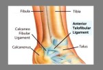
There are a variety of anatomical structures that make up the anatomy of the foot and ankle (Figure 1) including bones, joints, ligaments, muscles, tendons, and nerves. These will be reviewed in the sections of this chapter.
The ankle joint is formed by the connection of three bones. The ankle bone is called the talus. The top of the talus fits inside a socket that is formed by the lower end of the tibia (shinbone) and the fibula (the small bone of the lower leg).
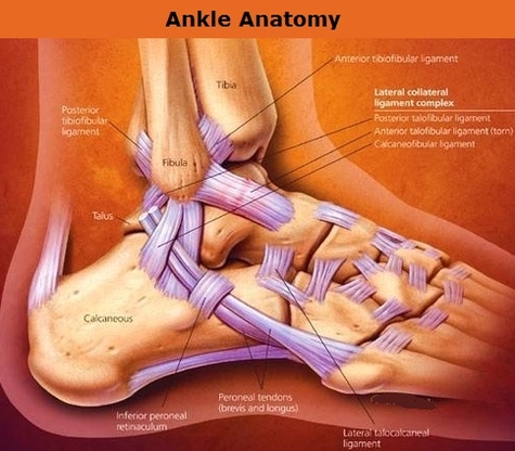
[edit on Wikidata] The ankle, or the talocrural region, is the region where the foot and the leg meet. The ankle includes three joints: the ankle joint proper or talocrural joint, the subtalar joint, and the inferior tibiofibular joint. The movements produced at this joint are dorsiflexion and plantarflexion of the foot.
ankle anatomy
Ankle anatomy
Post navigation
Previous Post
Next Post
Ankle ligaments