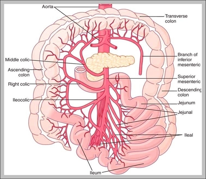
Blood Supply and Innervation of the Small Intestine. The neurovascular supply of any area of the body is the network in which the blood is pumped and the structures are…
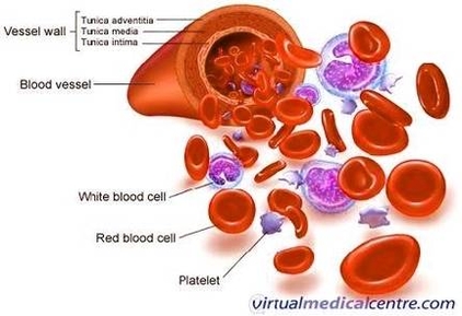
Blood Vessels. Blood vessels are the body’s highways that allow blood to flow quickly and efficiently from the heart to every region of the body and back again. The size…
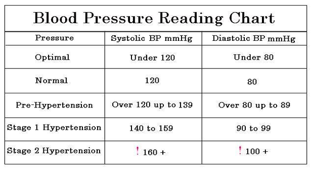
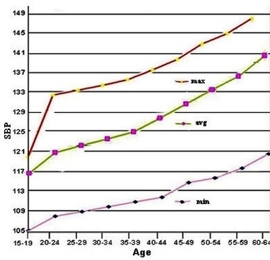
Blood pressure chart: What your reading means. By Mayo Clinic Staff. This blood pressure chart can help you figure out if your blood pressure is at a healthy level or…
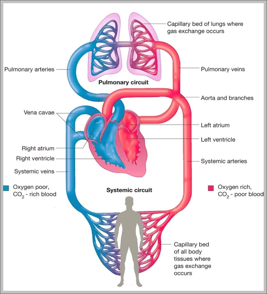
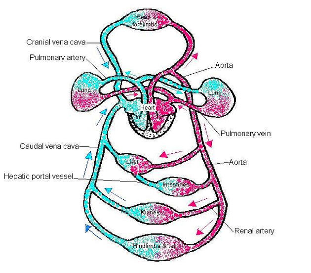
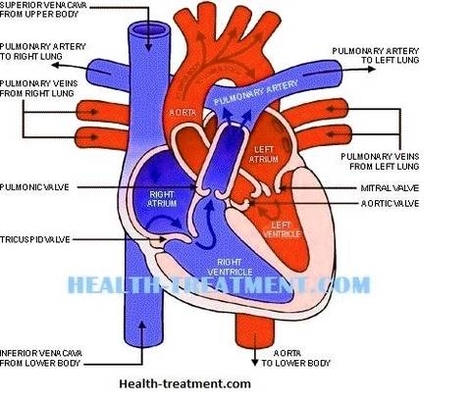
What is a Circulatory System Diagram. Systemic Circulation: After receiving oxygenated blood from the lungs the arteries of the systemic circulation system take the oxygenated blood from the heart to…
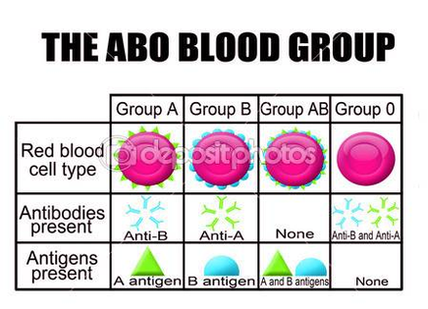
• Other gene which plays an important role in the determination of ABO blood groups is H gene with locus on chromosome 19. • Each individual inherits two ABO genes…
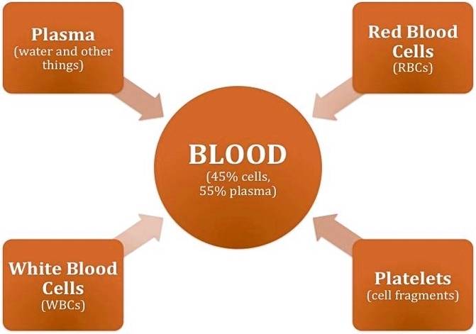
What is Blood. Blood is essential to life. Blood circulates through our body and delivers essential substances like oxygen and nutrients to the body’s cells. It also transports metabolic waste…
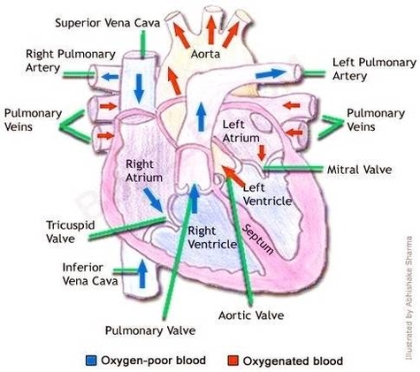
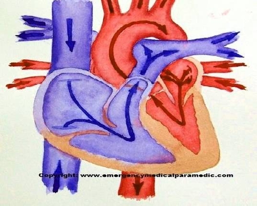
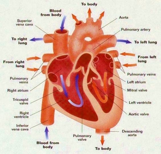
How Does the Blood Flow Through Your Heart 1 Right Side. Blood flows from your right atrium into your right ventricle through... 2 Left Side. Blood flows from your left…
How Does the Blood Flow Through Your Heart 1 Right Side. Blood flows from your right atrium into your right ventricle through... 2 Left Side. Blood flows from your left…
How Does the Blood Flow Through Your Heart 1 Right Side. Blood flows from your right atrium into your right ventricle through... 2 Left Side. Blood flows from your left…
What is a Circulatory System Diagram. Systemic Circulation: After receiving oxygenated blood from the lungs the arteries of the systemic circulation system take the oxygenated blood from the heart to…
A series of related blood types constitutes a blood group system, such as the Rh or ABO system. The frequencies of the ABO and Rh blood types vary from population…
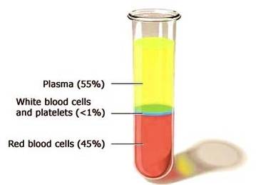
Composition of the Blood. The light yellow colored liquid on the top is the plasma, which accounts for about 55 percent of the blood volume and red blood cells is…