Internal Structure Of A Virus

Internal Structure of a Virus

A virus is a microscopic infectious particle that can reproduce only by infecting a host cell. Viruses are not considered living as they can’t reproduce by themselves. They are much smaller than the cells of living things and are basically just packages of nucleic acid and protein.

Basic Components

A typical virus consists of two primary components:

1. Nucleic Acid Genome: This is the genetic material of the virus, which can be either DNA or RNA.

2. Protein Capsid: This is a protective protein shell that encases the nucleic acid genome. The capsid is made from proteins encoded by viral genes within their genome. The shape of the capsid varies among different types of viruses and serves as one basis for their classification.

Many animal viruses also contain a lipid envelope along with some additional parts such as the neck, tail sheath, tail fibers, pins, and endplate to form a complete virion.

Capsid Shapes

Capsids can have different shapes:

1. Helical Capsids: These are made up of a single type of protein subunit stacked around a central axis to form a helical structure. This arrangement results in rod-shaped or filamentous virions. An example of a helical virus is the tobacco mosaic virus.

2. Icosahedral Capsids: These give viruses a spherical appearance at low magnification, but the protein subunits are actually arranged in a regular geometrical pattern.

Envelope
ome viruses have an envelope of phospholipids and proteins. The envelope is made from portions of the host’s cell membrane. It surrounds the capsid and helps protect the virus from the host’s immune system. The envelope may also have receptor molecules that can bind with host cells, making it easier for the virus to infect the cells.

Reproduction

Viruses reproduce by infecting their host cells and reprogramming them to become virus-making “factories”. They “commandeer” the host cell and use its resources to make more viruses.

Diversity

Viruses are very diverse. They come in different shapes and structures, have different kinds of genomes, and infect different hosts. Scientists estimate that there are roughly 10^31 viruses at any given moment.

In conclusion, viruses are tiny, non-living infectious particles with a simple structure. Despite their simplicity, they have a significant impact on all forms of life.

Internal Structure Of A Virus Diagram - Internal Structure Of A Virus Chart - Human anatomy diagrams and charts explained. This anatomy system diagram depicts Internal Structure Of A Virus with parts and labels. Best diagram to help learn about health, human body and medicine.

Internal Structure Of A Virus

Posterior View Of Internal Organs

The human body is a complex system of organs, each with its unique function. When viewed from the posterior (back), several key organs are visible. Here’s an overview of these organs and their functions:

1. Brain: The brain is the body’s control center. It forms the core of the central nervous system by creating, sending, and processing nerve impulses, thoughts, emotions, physical sensations, and more. The major areas of the brain include:
– The medulla: Helps control heart and lung function.
– The pons: Helps control eye and facial movement.
– The parietal lobe: Supports identifying objects and doing spatial reasoning.
– The frontal lobe: Plays a role in many conscious functions, including personality and movement.
– The occipital lobes: Primarily interprets vision signals.
– The temporal lobes: Play a role in numerous functions, including speech, scent recognition, and short-term memory.

2. Heart: The heart is the most important organ of the circulatory system, which helps deliver blood to the body. It has four chambers: two upper chambers called atria, and two lower chambers called ventricles. Blood flows into the right atrium from the veins of the heart and body (except the lungs) and then flows into the right ventricle. From there, it flows into the pulmonary artery, which has branches that reach the lungs. The lungs then oxygenate the blood.

3. Lungs: The lungs are vital for respiration. They oxygenate the blood and remove carbon dioxide, a waste product of metabolism. In the case of the paired lungs, a person can live without one of the pair.

4. Liver: The liver is a large organ that sits on the right side of the belly. It detoxifies chemicals and metabolizes drugs. The liver also makes proteins important for blood clotting and other functions.

5. Kidneys: The kidneys are two bean-shaped organs, each about the size of a fist. They are located just below the rib cage, one on each side of the spine. The kidneys filter out wastes, extra water, and salt through the urine. In the case of the paired kidneys, a person can live without one of the pair.

These organs are vital for survival, and any problem with these organs can quickly become life-threatening. The posterior view of the human body also includes the spine, which provides structural support and houses the spinal cord, an extension of the brain that communicates with the rest of the body. The posterior view also shows the muscles and bones that support these organs and enable movement.

In conclusion, the posterior view of the human body reveals a complex interplay of organs, each with its unique function, working together to maintain the body’s overall health and vitality..

Posterior View Of Internal Organs Diagram - Posterior View Of Internal Organs Chart - Human anatomy diagrams and charts explained. This anatomy system diagram depicts Posterior View Of Internal Organs with parts and labels. Best diagram to help learn about health, human body and medicine.

Posterior View Of Internal Organs

Internal Structure Of Long Bone

Internal Structure of Long Bones

Long bones, as the name suggests, are longer than they are wide. They are one of the types of bones classified based on their shape, and they play a crucial role in the skeletal system. Examples of long bones include the femur, tibia, fibula, humerus, ulna, and radius.

A long bone is primarily composed of two parts: the diaphysis and the epiphysis.

1. Diaphysis: The diaphysis, or the shaft, is the long, hollow, tubular structure that runs between the proximal and distal ends of the bone. The diaphysis is composed of a thick layer of compact bone, a dense and hard form of osseous tissue. Inside the diaphysis is the medullary cavity, which is filled with yellow bone marrow in an adult.

2. Epiphysis: The wider section at each end of the bone is called the epiphysis. Internally, the epiphysis is filled with spongy bone, another type of osseous tissue. Red bone marrow fills the spaces between the spongy bone in some long bones. Each epiphysis meets the diaphysis at the metaphysis.

3. Metaphysis: The metaphysis is the narrow area that contains the epiphyseal plate (growth plate), a layer of hyaline (transparent) cartilage in a growing bone. When the bone stops growing in early adulthood (approximately 18–21 years), the cartilage is replaced by osseous tissue and the epiphyseal plate becomes an epiphyseal line.

4. Endosteum: The medullary cavity has a delicate membranous lining called the endosteum. This is where bone growth, repair, and remodeling occur.

5. Periosteum: The outer surface of the bone is covered with a fibrous membrane called the periosteum. The periosteum contains blood vessels, nerves, and lymphatic vessels that nourish compact bone. Tendons and ligaments also attach to bones at the periosteum.

6. Articular Cartilage: In the region where the epiphyses meet other bones to form joints, the epiphyses are covered with articular cartilage, a thin layer of hyaline cartilage that reduces friction and acts as a shock absorber.

The internal structure of long bones is a marvel of nature’s engineering, providing strength, flexibility, and the ability to withstand stress. The intricate design of these bones allows them to perform their functions effectively, contributing to our ability to move and interact with our environment..

Internal Structure Of Long Bone Diagram - Internal Structure Of Long Bone Chart - Human anatomy diagrams and charts explained. This anatomy system diagram depicts Internal Structure Of Long Bone with parts and labels. Best diagram to help learn about health, human body and medicine.

Internal Structure Of Long Bone

Bird Internal Anatomy Diagram

Bird Internal Anatomy

Birds, as a group of vertebrates, exhibit a unique set of anatomical features, many of which are adaptations to facilitate flight.

1. Skeletal System: Birds have a lightweight skeletal system, with many bones being hollow (pneumatized) for structural strength. The number of hollow bones varies among species, with large gliding and soaring birds tending to have the most. The bird skeleton is highly adapted for flight, being lightweight but strong enough to withstand the stresses of taking off, flying, and landing.

2. Respiratory System: A bird’s respiratory system is much more efficient than that of mammals. Birds do not have a diaphragm like mammals, which mammals use to increase and decrease their chest cavity. Respiratory air sacs often form air pockets within the semi-hollow bones of the bird’s skeleton.

3. Digestive System: The development of a beak has led to the evolution of a specially adapted digestive system. Birds also lack teeth or even a true jaw, and instead have a beak, which is far more lightweight.

4. Vertebral Column: The vertebral column is divided into five sections of vertebrae: cervical, thoracic/dorsal, synsacrum, caudal, and pygostyle. The cervical vertebrae provide structural support to the neck and can number between 8 and as many as 25 vertebrae in certain swan species and other long-necked birds.

5. External Anatomy: The external anatomy of a bird includes features such as the beak, head, iris, pupil, mantle, lesser coverts, scapulars, coverts, tertials, rump, primaries, vent, thigh, tibio-tarsal articulation, tarsus, feet, tibia, belly, flanks, breast, throat, and wattle.

6. Rise of Nationalism: The Western concept of nationalism disrupted the Ottoman Empire’s traditional millet system. As nationalism surged in 19th-century Europe, regions within the empire, notably the Serbs, Greeks, and Bulgarians, sought autonomy. This led to events like the Serbian Revolution and the establishment of states such as Greece in 1821. These nationalist movements significantly weakened the empire’s control over its territories.

In conclusion, the internal anatomy of birds is a fascinating study of evolutionary adaptations that have enabled these creatures to conquer the skies. From their lightweight skeletal structure to their efficient respiratory system, each aspect of their anatomy plays a crucial role in

Bird Internal Anatomy Diagram Diagram - Bird Internal Anatomy Diagram Chart - Human anatomy diagrams and charts explained. This anatomy system diagram depicts Bird Internal Anatomy Diagram with parts and labels. Best diagram to help learn about health, human body and medicine.

Bird Internal Anatomy Diagram

Female Abdominal Anatomy And Internal Organs

The female abdominal anatomy is a complex and intricate system that houses various organs, each with its unique function. These organs are protected by the abdominal muscles, which include the rectus abdominis in front, the external obliques at the sides, and the latissimus dorsi muscles in the back.

Major Organs

1. Stomach, Small Intestine, and Large Intestine: These organs are responsible for digestion. They turn nutrients into usable energy and help dispose of solid waste.

2. Liver: Located in the upper right-hand part of the abdominal cavity, under the ribs, the liver processes blood, separating waste from nutrients.

3. Gallbladder: This tiny sack under the liver holds extra bile made by the liver until it is pumped into the small intestine. Bile helps break down fat.

4. Pancreas: This gland produces enzymes to help your body digest proteins, carbohydrates, and fats. It also makes hormones that help regulate the distribution of nutrients, including sugar.

5. Kidneys: Located near the back of the body, under the ribs, on each side of the spine, kidneys filter waste out of the bloodstream, which is passed out of the body as urine. They also help regulate levels of electrolytes, like salt and potassium, and produce certain hormones.

6. Suprarenal (Adrenal) Glands: These glands synthesize and secrete hormones that help the kidneys conserve sodium, thus conserving water. They also play a role in supporting the body’s sexual functions.

Female Reproductive Organs

1. Uterus (Womb): A hollow, pear-shaped organ located in a woman’s lower abdomen, between the bladder and the rectum.

2. Ovaries: Two female reproductive organs located in the pelvis.

3. Fallopian Tubes: These carry eggs from the ovaries to the uterus.

4. Cervix: The lower part of the uterus that opens into the vagina.

5. Vagina: The canal that joins the cervix to the outside of the body.

External Female Anatomy

1. Mons Pubis: The rounded, fleshy area on the front of the pelvic bone where pubic hair usually grows.

2. Labia Majora and Minora: The fleshy outer and inner folds of protective skin located on each side of the vaginal opening.

3. Clitoris: Sits at the top of the vulva, above the urethral opening.

4. Urethral Opening: The tube that carries urine from the bladder to the outside of the body.

These organs and structures work together to perform a variety of functions, including digestion, waste removal, reproduction, and hormone regulation. Understanding the female abdominal anatomy is crucial for maintaining health and diagnosing potential health issues..

Female Abdominal Anatomy And Internal Organs Diagram - Female Abdominal Anatomy And Internal Organs Chart - Human anatomy diagrams and charts explained. This anatomy system diagram depicts Female Abdominal Anatomy And Internal Organs with parts and labels. Best diagram to help learn about health, human body and medicine.

Female Abdominal Anatomy And Internal Organs

Internal Structure Of Human Stomach Visual

The human stomach, a key organ in the digestive system, is a complex structure with several distinct regions and layers. It plays a crucial role in the digestion of food and absorption of nutrients.

Location and Structure

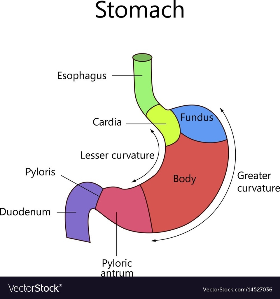
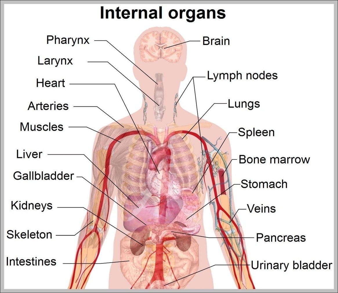
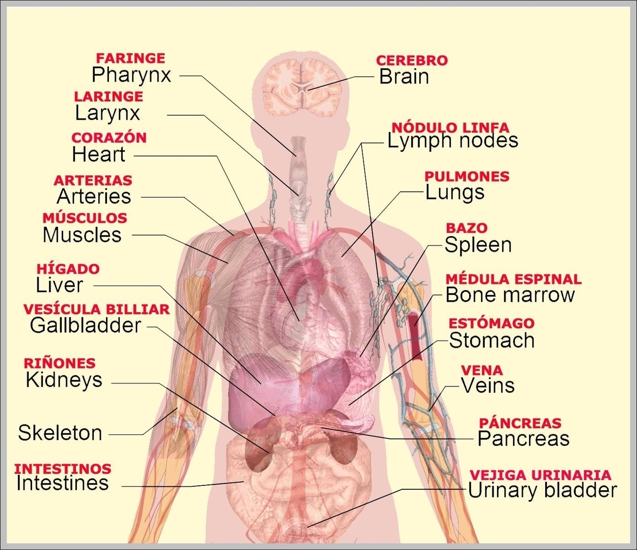
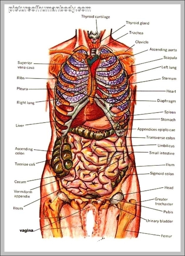
The stomach is located in the upper abdomen on the left side of the body. It is a J-shaped organ that spans the region between the cardiac and pyloric orifices of the gastrointestinal tract. The stomach’s convex lateral surface is known as the greater curvature, while the concave medial border is the lesser curvature.

Parts of the Stomach

The stomach comprises four major regions:
1. Cardia: The area around the opening where the esophagus connects to the stomach.
2. Fundus: The dome-shaped part located to the left of the cardia.
3. Body: The main, central region of the stomach.
4. Pylorus: The lower part of the stomach that connects to the duodenum.

Layers of the Stomach

The stomach wall consists of several layers:
1. Mucosa: The innermost layer, which produces enzymes and acids for digestion.
2. Submucosa: Contains connective tissue, blood vessels, lymph vessels, and nerve cells.
3. Muscularis Externa: The primary muscle of the stomach, responsible for churning and mixing food.
4. Serosa: The outermost layer, a membrane that covers the stomach.

Function of the Stomach

The stomach’s primary function is to digest food and send it to the small intestine. It temporarily stores food, contracts and relaxes to mix and break down food, and produces enzymes and other specialized cells to digest food. The stomach works in conjunction with the rest of the gastrointestinal tract to break down food and liquid, absorb nutrients and water, and expel waste products of digestion through the large intestine.

Blood Supply and Innervation

The stomach receives its blood supply mainly from the celiac trunk. Innervation is provided via the vagus nerves and the celiac plexus.

Microscopic Anatomy

The inner part of the stomach lining, the gastric mucosa, consists of an outer layer of column-shaped cells, a lamina propria, and a thin layer of smooth muscle called the muscularis mucosa. Beneath the mucosa lies the submucosa, consisting of fibrous connective tissue.

In conclusion, the human stomach is a complex organ with a detailed internal structure. Its various parts and layers work together to perform the essential function of digesting food and absorbing nutrients, making it a vital component of the human digestive system..

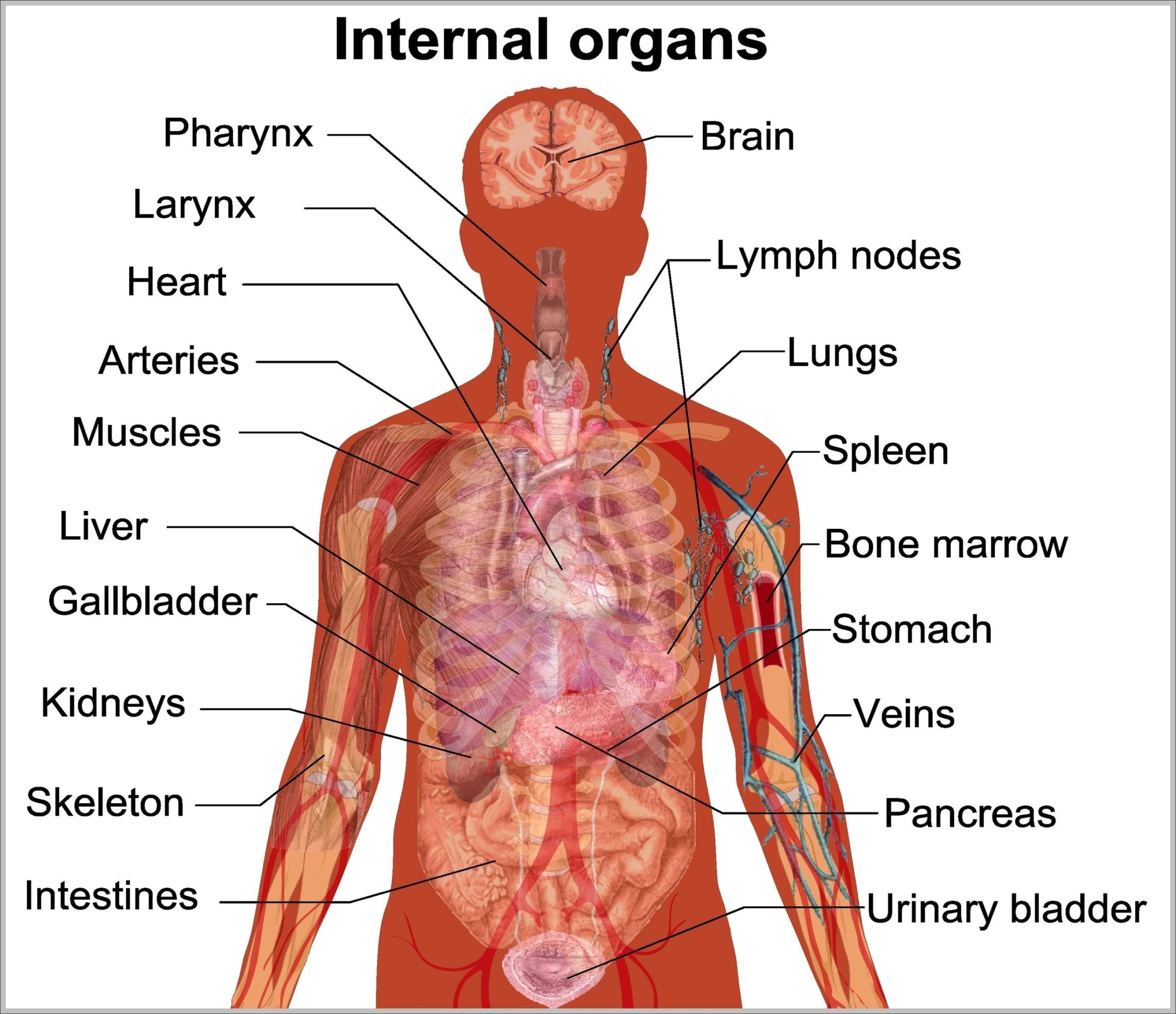
Internal Structure Of Human Stomach Visual Diagram - Internal Structure Of Human Stomach Visual Chart - Human anatomy diagrams and charts explained. This anatomy system diagram depicts Internal Structure Of Human Stomach Visual with parts and labels. Best diagram to help learn about health, human body and medicine.

Internal Structure Of Human Stomach Visual

Internal Body Anatomy Image

Internal Body Anatomy Image Diagram - Internal Body Anatomy Image Chart - Human anatomy diagrams and charts explained. This anatomy system diagram depicts Internal Body Anatomy Image with parts and labels. Best diagram to help learn about health, human body and medicine.

Internal Body Anatomy Image

Human Anatomy Internal Organs Image

Human internal organs. Stomach and lungs, kidneys and heart, brain and liver. Body parts vector anatomy set. Illustration health organ collection for system Human internal organs. Stomach and lungs, kidneys and heart, brain and liver.
10,426 female anatomy internal organs stock photos and images available, or start a new search to explore more stock photos and images. Human body internal organs, skeleton, skeletal bones,… Human anatomy infographic elements with set of internal organs isolated on white background and placed in female body.

Human Anatomy Internal Organs Image Diagram - Human Anatomy Internal Organs Image Chart - Human anatomy diagrams and charts explained. This anatomy system diagram depicts Human Anatomy Internal Organs Image with parts and labels. Best diagram to help learn about health, human body and medicine.

Human Anatomy Internal Organs Image

Human Body Internal Organs Diagram Image

7,751 organs of the human body diagram stock illustrations and vector graphics available royalty-free, or start a new search to explore more great stock images and vector art. internal organs and circulatory system Vector isolated illustration of human internal organs and circulatory system in man body.
Human internal organs vector Vector isolated illustration of human internal organs in male body. Stomach, liver, intestine, bladder, lung, testicle, uterus, spine, pancreas, kidney, heart, bladder icon. Donor medical poster organs of the human body diagram stock illustrations

Human Body Internal Organs Diagram Image Diagram - Human Body Internal Organs Diagram Image Chart - Human anatomy diagrams and charts explained. This anatomy system diagram depicts Human Body Internal Organs Diagram Image with parts and labels. Best diagram to help learn about health, human body and medicine.

Human Body Internal Organs Diagram Image

Human Anatomy Diagram Internal Organs Image

Human internal organs vector Vector isolated illustration of human internal organs in male body. Stomach, liver, intestine, bladder, lung, testicle, uterus, spine, pancreas, kidney, heart, bladder icon. Donor medical poster organs of the human body diagram stock illustrations
7,751 organs of the human body diagram stock illustrations and vector graphics available royalty-free, or start a new search to explore more great stock images and vector art. internal organs and circulatory system Vector isolated illustration of human internal organs and circulatory system in man body.

Human Anatomy Diagram Internal Organs Image Diagram - Human Anatomy Diagram Internal Organs Image Chart - Human anatomy diagrams and charts explained. This anatomy system diagram depicts Human Anatomy Diagram Internal Organs Image with parts and labels. Best diagram to help learn about health, human body and medicine.

Human Anatomy Diagram Internal Organs Image

Internal Organs Pictures Image

407,393 human internal organ stock photos and images available, or search for human internal organ icons or human internal organ illustrations to find more great stock photos and pictures. Organs, anatomy flat line icons set. Human bones, stomach, brain, heart, bladder, nervous system vector illustrations.
Medicine, internal organs. 3d vector icon set human internal organ stock illustrations Human heart, brain, eye, tooth, lungs, liver, stomach, kidney,… 3d illustration of human body organs The human body is the entire structure of a human being.

Internal Organs Pictures Image Diagram - Internal Organs Pictures Image Chart - Human anatomy diagrams and charts explained. This anatomy system diagram depicts Internal Organs Pictures Image with parts and labels. Best diagram to help learn about health, human body and medicine.

Internal Organs Pictures Image

Human Internal Organ Diagram Image

80,662 human internal organ stock photos and images available, or search for human internal organ icons or human internal organ illustrations to find more great stock photos and pictures.
7,751 organs of the human body diagram stock illustrations and vector graphics available royalty-free, or start a new search to explore more great stock images and vector art. internal organs and circulatory system Vector isolated illustration of human internal organs and circulatory system in man body.

Human Internal Organ Diagram Image Diagram - Human Internal Organ Diagram Image Chart - Human anatomy diagrams and charts explained. This anatomy system diagram depicts Human Internal Organ Diagram Image with parts and labels. Best diagram to help learn about health, human body and medicine.

Human Internal Organ Diagram Image scaled

Human Body Internal Organs Image

Medicine, internal organs. 3d vector icon set human internal organ stock illustrations Human heart, brain, eye, tooth, lungs, liver, stomach, kidney,… 3d illustration of human body organs The human body is the entire structure of a human being.
407,393 human internal organ stock photos and images available, or search for human internal organ icons or human internal organ illustrations to find more great stock photos and pictures. Organs, anatomy flat line icons set. Human bones, stomach, brain, heart, bladder, nervous system vector illustrations.

Human Body Internal Organs Image Diagram - Human Body Internal Organs Image Chart - Human anatomy diagrams and charts explained. This anatomy system diagram depicts Human Body Internal Organs Image with parts and labels. Best diagram to help learn about health, human body and medicine.

Human Body Internal Organs Image

Human Anatomy Chart Internal Organs Image

80,933 human internal organ stock photos and images available or search for human internal organ icons or human internal organ illustrations to find more great stock photos and pictures.
Human internal organs vector Vector isolated illustration of human internal organs in male body. Stomach, liver, intestine, bladder, lung, testicle, uterus, spine, pancreas, kidney, heart, bladder icon. Donor medical poster organs of the human body diagram stock illustrations

Human Anatomy Chart Internal Organs Image Diagram - Human Anatomy Chart Internal Organs Image Chart - Human anatomy diagrams and charts explained. This anatomy system diagram depicts Human Anatomy Chart Internal Organs Image with parts and labels. Best diagram to help learn about health, human body and medicine.

Human Anatomy Chart Internal Organs Image

Internal Organs Anatomy Image

Internal Organs Anatomy Image Diagram - Internal Organs Anatomy Image Chart - Human anatomy diagrams and charts explained. This anatomy system diagram depicts Internal Organs Anatomy Image with parts and labels. Best diagram to help learn about health, human body and medicine.

Internal Organs Anatomy Image

Internal Organ Chart Image

An organ is a collection of tissues that have a specific role to play in the human body. They also work in tandem to form organ systems, like the digestive system or the circulatory system. Here is some information about the most important internal organs of the human body, beginning with the head, and continuing downward.
7,751 organs of the human body diagram stock illustrations and vector graphics available royalty-free, or start a new search to explore more great stock images and vector art. internal organs and circulatory system Vector isolated illustration of human internal organs and circulatory system in man body.

Internal Organ Chart Image Diagram - Internal Organ Chart Image Chart - Human anatomy diagrams and charts explained. This anatomy system diagram depicts Internal Organ Chart Image with parts and labels. Best diagram to help learn about health, human body and medicine.

Internal Organ Chart Image scaled

Human Internal Organs Pictures Image

407,393 human internal organ stock photos and images available, or search for human internal organ icons or human internal organ illustrations to find more great stock photos and pictures. Organs, anatomy flat line icons set. Human bones, stomach, brain, heart, bladder, nervous system vector illustrations.
Medicine, internal organs. 3d vector icon set human internal organ stock illustrations Human heart, brain, eye, tooth, lungs, liver, stomach, kidney,… 3d illustration of human body organs The human body is the entire structure of a human being.

Human Internal Organs Pictures Image Diagram - Human Internal Organs Pictures Image Chart - Human anatomy diagrams and charts explained. This anatomy system diagram depicts Human Internal Organs Pictures Image with parts and labels. Best diagram to help learn about health, human body and medicine.

Human Internal Organs Pictures Image scaled

Picture Of The Internal Human Body Image

86,960 internal organs of the human body pics stock photos and images available, or start a new search to explore more stock photos and images. 3D illustration of Heart, medical concept.
Body, internal parts of body, brain, bladder, liver, stomach, kidneys, locomotor system, intestinal tract, pancreas Human Organs The human body is the entire structure of a human being and comprises a head, neck, trunk (which includes the thorax and abdomen), two arms and hands and two legs and feet.

Picture Of The Internal Human Body Image Diagram - Picture Of The Internal Human Body Image Chart - Human anatomy diagrams and charts explained. This anatomy system diagram depicts Picture Of The Internal Human Body Image with parts and labels. Best diagram to help learn about health, human body and medicine.

Picture Of The Internal Human Body Image

Internal Abdominal Oblique Image

The abdominal internal oblique muscle, also internal oblique muscle or interior oblique, is an abdominal muscle in the abdominal wall that lies below the external oblique muscle and just above the transverse abdominal muscle . The abdominal internal oblique muscle. Muscles of the trunk.
(T12), Iliohypogastric n. (L1) and Ilioinguinal n. (L1) The abdominal internal oblique muscle, also internal oblique muscle or interior oblique, is an abdominal muscle in the abdominal wall that lies below the external oblique muscle and just above the transverse abdominal muscle .

Internal Abdominal Oblique Image Diagram - Internal Abdominal Oblique Image Chart - Human anatomy diagrams and charts explained. This anatomy system diagram depicts Internal Abdominal Oblique Image with parts and labels. Best diagram to help learn about health, human body and medicine.

Internal Abdominal Oblique Image

Picture Internal Organs Image

Browse 407,393 human internal organ stock photos and images available, or search for human internal organ icons or human internal organ illustrations to find more great stock photos and pictures. Organs, anatomy flat line icons set. Human bones, stomach, brain, heart, bladder, nervous system vector illustrations.
Medicine, internal organs. 3d vector icon set human internal organ stock illustrations Human heart, brain, eye, tooth, lungs, liver, stomach, kidney,… 3d illustration of human body organs The human body is the entire structure of a human being.
Vector icons of human organs: pancreas, brain, heart, liver, lungs, spleen, intestines, kidneys,and stomach. Human man people anatomy internal organs system banner poster scheme. Medicine education concept. Vector flat cartoon isolated graphic design illustration Human man people anatomy internal organs system banner poster scheme.

Picture Internal Organs Image Diagram - Picture Internal Organs Image Chart - Human anatomy diagrams and charts explained. This anatomy system diagram depicts Picture Internal Organs Image with parts and labels. Best diagram to help learn about health, human body and medicine.

Picture Internal Organs Image scaled