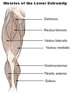
Lower Extremity Muscles Diagram
The primary muscle in this part of the body is the gastrocnemius, which gives the calf its signature bulging, muscular appearance. The anterior tibial, posterior tibial and fibular arteries are responsible for blood supply to the lower leg. The lower leg makes up a large portion of an individual’s overall body weight.
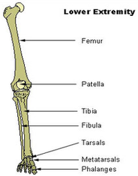
Key facts about the lower extremity Hip and pelvis Bones: hip bones, saccrum, coccyx Hip jo … Thigh Bones: femur Joints: hip and knee Muscle … Knee Bones: tibia, fibula, patella Type: hing … Leg Bones: tibia, fibula Joints: knee and an … Ankle and foot Ankle joint: hinged joint capable of …
The leg muscles and tendons produce tension, stabilize the joints of the legs, and create movement. The main muscle groups in the legs are: quadriceps, hamstrings, adductors in the upper leg or thigh, and the calves in the lower legs.