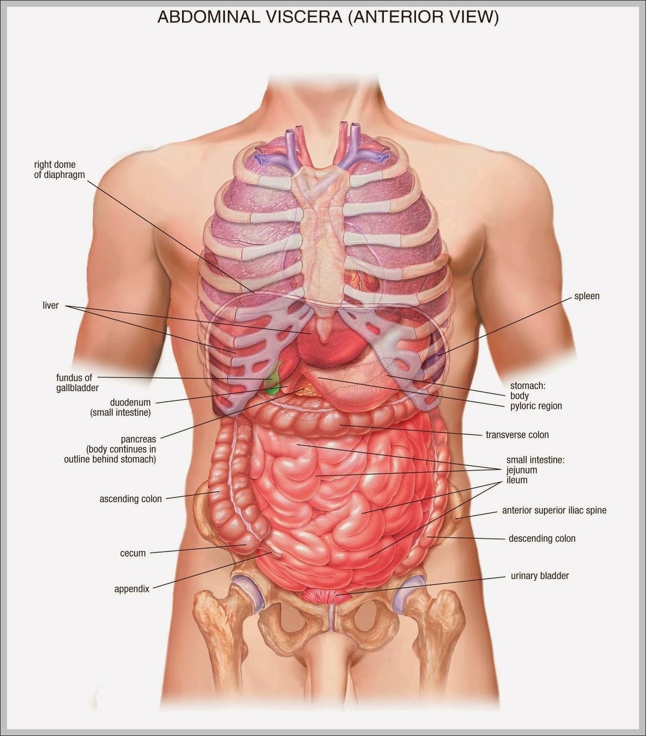
Human Abdomen Organs With Highlighted Stomach Examined
The human abdomen is a complex structure housing several vital organs. Let’s examine these organs, with a special focus on the stomach.
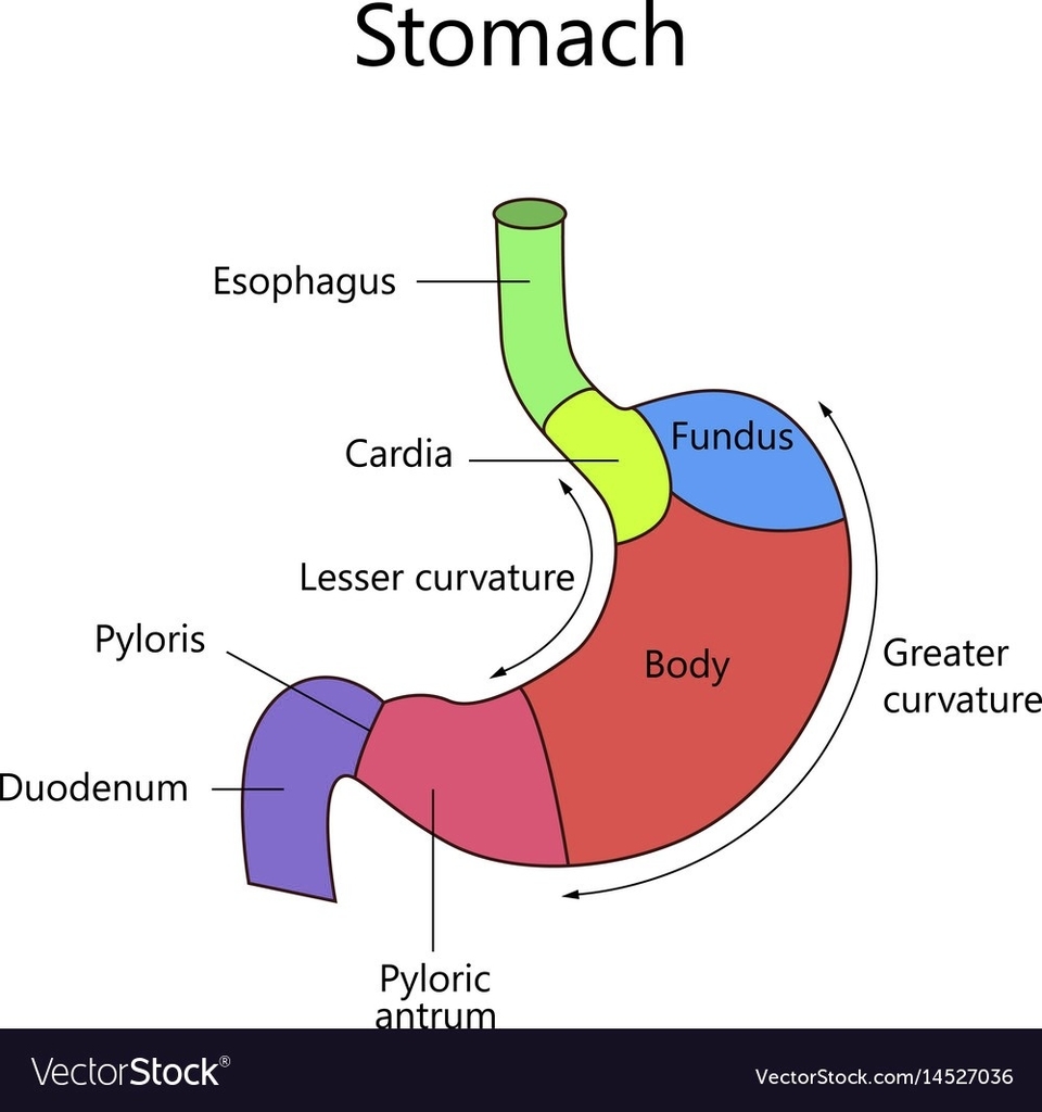
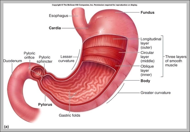
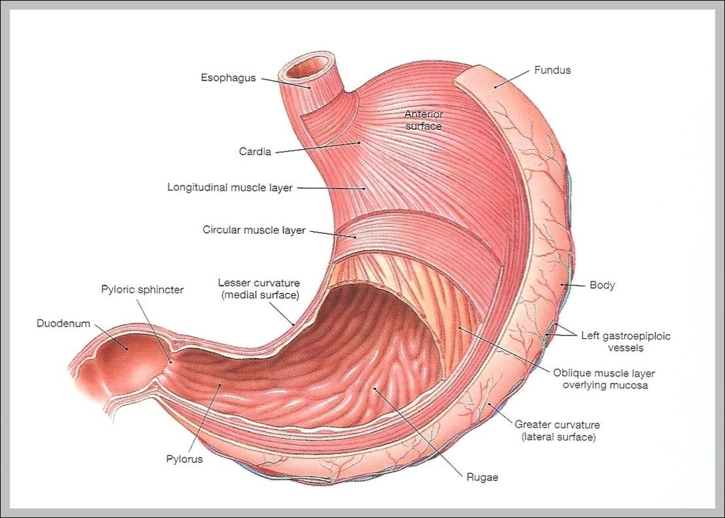
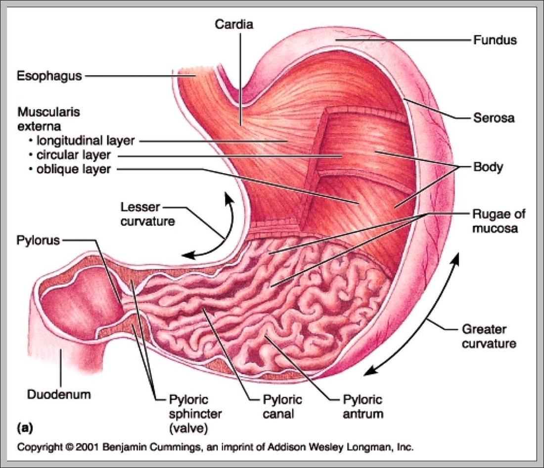
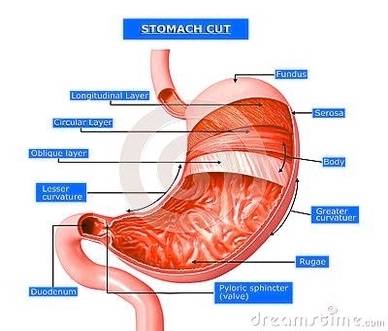
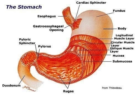
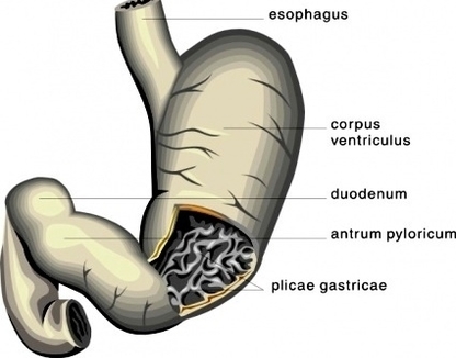
1. Stomach: Located in the upper part of the abdomen, the stomach plays a crucial role in digestion. It receives food from the esophagus and breaks it down both mechanically and chemically. The stomach’s three layers – the oblique layer, the middle circular layer, and the external longitudinal layer – work together to churn food. This mechanical breakdown is complemented by chemical digestion through stomach acids, including hydrochloric acid. The stomach also stores food until it’s ready to move further along the digestive tract.
2. Liver: Situated at the top of the abdominal cavity, the liver is the body’s largest organ. It acts as a filtration system, eliminating toxins and producing bile, which aids in the digestion and absorption of fats and fat-soluble vitamins.
3. Gallbladder: This small sac beneath the liver stores extra bile produced by the liver until it’s needed in the small intestine. Bile is crucial for digesting fats, excreting cholesterol, and even has antimicrobial activity.
4. Pancreas: This gland produces enzymes that help your body digest proteins, carbohydrates, and fats. It also makes hormones that help regulate the distribution of nutrients, including sugar.
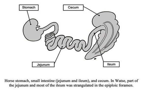
5. Small Intestine: Occupying most of the abdominal cavity, this 21-foot long tube is where the majority of digestion occurs. It breaks down fats, starches, and proteins into fatty acids, which can then be absorbed.
6. Large Intestine: Despite its name, the large intestine is shorter than the small intestine but larger in girth. It’s the last part of the digestive tract and is made up of the cecum, colon, and rectum.
7. Kidneys: Positioned behind the intestines, the kidneys contain an estimated 1 million filtering units called nephrons. They play a vital role in processing the blood before it goes into general circulation.
8. Adrenal Glands: Located on top of the kidneys, these glands synthesize and secrete different sets of hormones. These hormones help the kidneys conserve sodium and water, and also support the body’s sexual functions.
9. Ureters: These two tubes carry urine from the kidneys to the urinary bladder.
10. Ribs: The main bones in the abdominal region, the ribs protect vital internal organs.
In conclusion, the human abdomen is a marvel of biological engineering, with each organ playing a unique and vital role in maintaining the body’s overall health and functionality. The stomach, in particular, serves as a critical junction in the digestive process, preparing food for further digestion and absorption in the intestines..
Human Abdomen Organs With Highlighted Stomach Examined Diagram - Human Abdomen Organs With Highlighted Stomach Examined Chart - Human anatomy diagrams and charts explained. This anatomy system diagram depicts Human Abdomen Organs With Highlighted Stomach Examined with parts and labels. Best diagram to help learn about health, human body and medicine.