Human Anatomy For Muscle, Reproductive, And Skeleton

Human Anatomy: Muscle, Reproductive, and Skeletal Systems
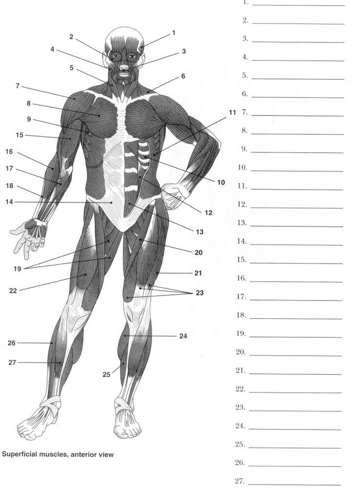
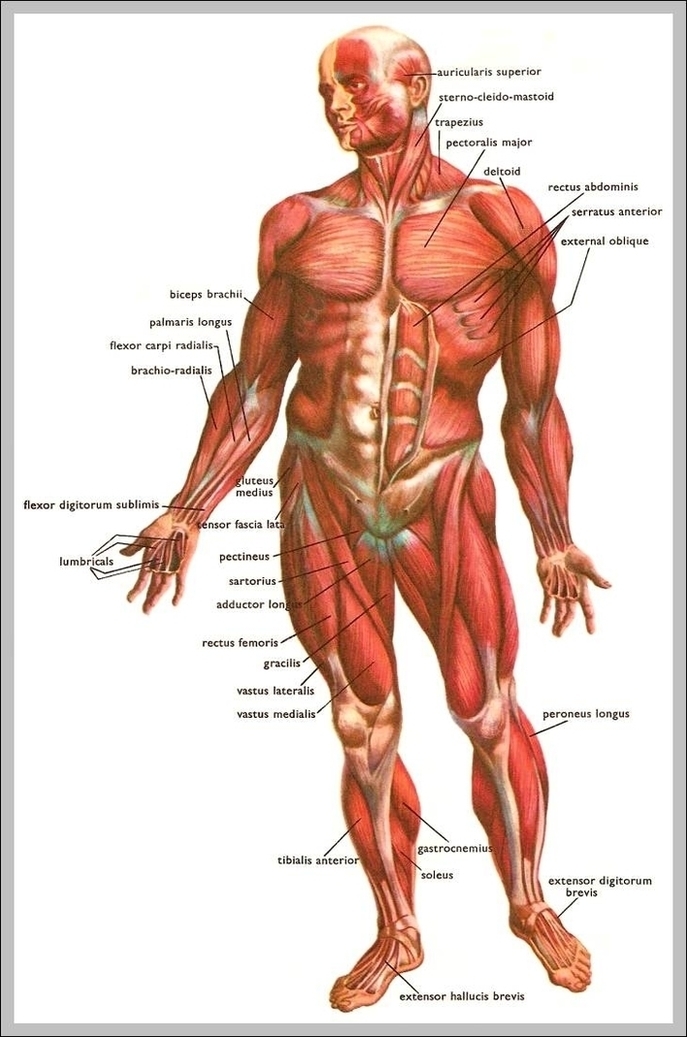
Muscular System
The muscular system is responsible for movement, posture, and balance. It consists of three types of muscles: skeletal, smooth, and cardiac.

– Skeletal Muscles: These muscles are attached to the bones by tendons and work in groups to move the skeleton. They make up about 40% of a person’s body weight.
– Smooth Muscles: Found in the walls of hollow organs, blood vessels, and respiratory passageways, they contract in response to stimuli and nerve impulses.
– Cardiac Muscles: These muscles make up the walls of the heart and are responsible for the rhythmic contractions that pump blood through the body.

Reproductive System
The reproductive system is responsible for human reproduction and bearing live offspring?. It includes both internal and external genitalia?.

– Male Reproductive System: The male reproductive system includes the testes that produce sperm and a penis for delivery?. The sperm mature in the testes and then enter the epididymis for further maturation?.
– Female Reproductive System: The female reproductive system includes the ovaries that produce eggs, a uterus for baby development, and breasts for milk production?. The ovum is released at a specific time in the reproductive cycle for internal fertilization by sperm cells?.
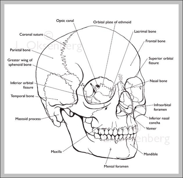
keletal System
The skeletal system serves as a framework for the body, providing shape, stability, and protection of internal organs. It consists of 206 bones, ligaments, and cartilages.

– Axial Skeleton: This includes the vertebral column (the spine) and much of the skull, providing the main support of the trunk.
– Appendicular Skeleton: This includes the pelvic (hip) and pectoral (shoulder) girdles and the bones and cartilages of the limbs.

In conclusion, the muscular, reproductive, and skeletal systems play crucial roles in the functioning of the human body. Each system has a unique structure and specific role, contributing to our movement, reproduction, and structural support.

Human Anatomy For Muscle, Reproductive, And Skeleton Diagram - Human Anatomy For Muscle, Reproductive, And Skeleton Chart - Human anatomy diagrams and charts explained. This anatomy system diagram depicts Human Anatomy For Muscle, Reproductive, And Skeleton with parts and labels. Best diagram to help learn about health, human body and medicine.

Human Anatomy For Muscle, Reproductive, And Skeleton

Human Leg Muscle Anatomy Examined

The human leg, a complex structure with numerous muscles, plays a pivotal role in body movement and support. The majority of leg muscles are considered long muscles, stretching great distances to move skeletal bones and facilitate body movement.

Upper Leg Muscles

The upper leg comprises the quadriceps and hamstrings. The quadriceps, the body’s strongest and leanest muscles, are major extensors of the knee. They include:

1. Vastus lateralis: The largest of the quadriceps, it extends from the top of the femur to the kneecap.
2. Vastus medialis: A teardrop-shaped muscle of the inner thigh, it attaches along the femur and down to the inner border of the kneecap.
3. Vastus intermedius: Located between the vastus medialis and the vastus lateralis at the front of the femur, it is the deepest of the quadriceps muscles.
4. Rectus femoris: This muscle attaches to the kneecap.

The hamstrings, three muscles at the back of the thigh, affect hip and knee movement. They include:

1. Biceps femoris: This long muscle flexes the knee.
2. Semimembranosus: This long muscle extends from the pelvis to the tibia.
3. Semitendinosus: This muscle extends the thigh and flexes the knee.

Lower Leg Muscles

The lower leg muscles, supported by the fibula and the tibia (shinbone), are pivotal to movement of the ankle, foot, and toes. Some of the major muscles of the calf include:

1. Gastrocnemius (calf muscle): One of the large muscles of the leg, it connects to the heel. It flexes and extends the foot, ankle, and knee.
2. Soleus: This muscle extends from the back of the knee to the heel. It is important in walking and standing.
3. Plantaris: This small, thin muscle is absent in about 10 percent of people. The gastrocnemius muscle supersedes its function.

The Achilles tendon, connecting the plantaris, gastrocnemius, and soleus muscles to the heel bone, stores the elastic energy needed for running, jumping, and other physical activity.

Functional Groups

The leg muscles are organized into three groups: anterior (dorsiflexor) group, posterior (plantar flexor) group, and lateral (fibular) group. These groups produce different movements in the ankle and foot, crucial for activities such as walking, running, and dancing.

The anterior group, including the tibialis anterior, extensor digitorum longus, fibularis tertius, and extensor hallucis longus, primarily produces dorsiflexion of the foot at the ankle joint. The posterior group, comprising the gastrocnemius, plantaris, soleus

Human Leg Muscle Anatomy Examined Diagram - Human Leg Muscle Anatomy Examined Chart - Human anatomy diagrams and charts explained. This anatomy system diagram depicts Human Leg Muscle Anatomy Examined with parts and labels. Best diagram to help learn about health, human body and medicine.

Human Leg Muscle Anatomy Examined

Human Earanatomy Of Human Ear

Anatomy of the Human Ear

The human ear is a complex organ that serves two primary functions: hearing and maintaining balance. It is anatomically divided into three parts: the outer ear, the middle ear, and the inner ear.

1. Outer Ear

The outer ear consists of the visible portion called the auricle or pinna, which projects from the side of the head, and the short external auditory canal. The inner end of this canal is closed by the tympanic membrane, commonly known as the eardrum. The function of the outer ear is to collect sound waves and guide them to the tympanic membrane.

2. Middle Ear

The middle ear is a narrow air-filled cavity in the temporal bone. It is spanned by a chain of three tiny bones — the malleus (hammer), incus (anvil), and stapes (stirrup), collectively known as the auditory ossicles. This ossicular chain conducts sound from the tympanic membrane to the inner ear.

3. Inner Ear

The inner ear, also known as the labyrinth, is a complicated system of fluid-filled passages and cavities located deep within the temporal bone. It consists of two functional units: the vestibular apparatus, which contains the sensory organs of postural equilibrium, and the cochlea, which contains the sensory organ of hearing. These sensory organs are highly specialized endings of the eighth cranial nerve, also known as the vestibulocochlear nerve.

Function of the Ear

The primary function of the ear is hearing, which involves the conversion of sound waves into electrochemical impulses, a process known as transduction. The ear also maintains the sense of balance or equilibrium. The vestibular apparatus provides information about the position and movements of the head, while the cochlea provides hearing information.

Clinical Relations

Various conditions can affect the ear, including otitis (inflammation of the ear), blockage of the auditory (Eustachian) tube, and high tone deafness. Understanding the anatomy of the ear is crucial for diagnosing and treating these conditions.

In conclusion, the human ear is a remarkable organ that plays a vital role in our ability to interact with our environment. Its complex structure enables it to perform its dual functions of hearing and maintaining balance, making it an essential part of our sensory system..

Human Earanatomy Of Human Ear Diagram - Human Earanatomy Of Human Ear Chart - Human anatomy diagrams and charts explained. This anatomy system diagram depicts Human Earanatomy Of Human Ear with parts and labels. Best diagram to help learn about health, human body and medicine.

Human Earanatomy Of Human Ear

External Anatomy Of The Left Human Eye

The human eye is a complex organ that allows us to perceive the world around us. The external anatomy of the eye includes several key components, each with a specific function.

1. Orbit: The eye sits in a protective bony socket called the orbit.

2. Extraocular Muscles: Six extraocular muscles in the orbit are attached to the eye. These muscles move the eye up and down, side to side, and rotate the eye.

3. Sclera: The extraocular muscles are attached to the white part of the eye called the sclera. This is a strong layer of tissue that covers nearly the entire surface of the eyeball.

4. Conjunctiva: The surface of the eye and the inner surface of the eyelids are covered with a clear membrane called the conjunctiva.

5. Tear Film: The layers of the tear film keep the front of the eye lubricated. Tears lubricate the eye and are made up of three layers. These three layers together are called the tear film.

6. Cornea: Light is focused into the eye through the clear, dome-shaped front portion of the eye called the cornea.

7. Anterior Chamber: Behind the cornea is a fluid-filled space called the anterior chamber. The fluid is called aqueous humor.

8. Iris and Pupil: Behind the anterior chamber is the eye’s iris (the colored part of the eye) and the dark hole in the middle called the pupil. Muscles in the iris dilate (widen) or constrict (narrow) the pupil to control the amount of light reaching the back of the eye.

9. Lens: Directly behind the pupil sits the lens. The lens focuses light toward the back of the eye. The lens changes shape to help the eye focus on objects up close.

10. Eyelids: The upper and lower eyelids form a moist region around the eye, and protect the surface of the eye from injury, infection, and disease.

11. Eyelashes: The eyelashes are the hairs that grow along the edges of the upper and lower eyelids. The eyelashes protect the eye from foreign particles, such as dust, pollen, and debris.

12. Meibomian Glands: Meibomian glands are the oil glands located inside the eyelids. Their opening pores line the edges of the eyelids, near the eyelashes.

Each of these components plays a crucial role in the overall function of the eye. They work together to protect the eye, control the amount of light that enters, and focus that light onto the retina, allowing us to see the world around us..

External Anatomy Of The Left Human Eye Diagram - External Anatomy Of The Left Human Eye Chart - Human anatomy diagrams and charts explained. This anatomy system diagram depicts External Anatomy Of The Left Human Eye with parts and labels. Best diagram to help learn about health, human body and medicine.

External Anatomy Of The Left Human Eye

Ear Anatomysimple Ear Anatomy Visual

Anatomy of the Human Ear

The human ear is a complex organ that serves two primary functions: hearing and maintaining balance. It is anatomically divided into three main parts: the outer ear, the middle ear, and the inner ear.

1. Outer Ear

The outer ear consists of the visible portion called the auricle, or pinna, and the external auditory canal. The auricle collects sound waves and guides them into the auditory canal. The inner end of the canal is closed by the tympanic membrane, commonly known as the eardrum. The function of the outer ear is to collect sound waves and guide them to the tympanic membrane.

2. Middle Ear

The middle ear is an air-filled cavity in the temporal bone. It contains a chain of three tiny bones — the malleus (hammer), incus (anvil), and stapes (stirrup), collectively known as the auditory ossicles. These bones conduct sound from the tympanic membrane to the inner ear.

3. Inner Ear

The inner ear, also known as the labyrinth, is a complex system of fluid-filled passages and cavities. It consists of two functional units: the vestibular apparatus and the cochlea. The vestibular apparatus, which includes the vestibule and semicircular canals, maintains balance. The cochlea, on the other hand, is responsible for hearing. These sensory organs are highly specialized endings of the eighth cranial nerve, also known as the vestibulocochlear nerve

Ear Anatomysimple Ear Anatomy Visual Diagram - Ear Anatomysimple Ear Anatomy Visual Chart - Human anatomy diagrams and charts explained. This anatomy system diagram depicts Ear Anatomysimple Ear Anatomy Visual with parts and labels. Best diagram to help learn about health, human body and medicine.

Ear Anatomysimple Ear Anatomy Visual

Brain Anatomy Diagram

Brain Anatomy

The brain, a complex organ, controls thought, memory, emotion, touch, motor skills, vision, breathing, temperature, hunger, and every process that regulates our body. It is primarily composed of nerve cells, also known as neurons. Blood vessels supply oxygen and nutrients to the neurons of the brain.

Composition

Weighing about 3 pounds in the average adult, the brain is about 60% fat. The remaining 40% is a combination of water, protein, carbohydrates, and salts. The brain itself is not a muscle. It contains blood vessels and nerves, including neurons and glial cells.

Gray Matter and White Matter

Gray and white matter are two different regions of the central nervous system. In the brain, gray matter refers to the darker, outer portion, while white matter describes the lighter, inner section underneath. Gray matter is primarily composed of neuron somas (the round central cell bodies), and white matter is mostly made of axons (the long stems that connect neurons together) wrapped in myelin (a protective coating). Gray matter is primarily responsible for processing and interpreting information, while white matter transmits that information to other parts of the nervous system.

Brain Function

The brain sends and receives chemical and electrical signals throughout the body. Different signals control different processes, and your brain interprets each. Some make you feel tired, for example, while others make you feel pain. Some messages are kept within the brain, while others are relayed through the spine and across the body’s vast network of nerves to distant extremities.

Main Parts of the Brain and Their Functions

At a high level, the brain can be divided into the cerebrum, brainstem, and cerebellum.

1. Cerebrum: The cerebrum (front of brain) comprises gray matter (the cerebral cortex) and white matter at its center. The largest part of the brain, the cerebrum initiates and coordinates movement and regulates temperature. Other areas of the cerebrum enable speech, judgment, thinking and reasoning, problem-solving, emotions, and learning. Other functions relate to vision, hearing, touch, and other senses.

2. Cerebral Cortex: The cortex has a large surface area due to its folds, and comprises about half of the brain’s weight. The cerebral cortex is divided into two halves, or hemispheres. It is covered with ridges (gyri) and folds (sulci).

3. Brain Stem: The brain stem controls the flow of messages between the brain and the rest of the body, and it also controls basic body functions such as breathing, swallowing, heart rate, blood pressure, consciousness, and whether one is awake or sleepy.

4. Cerebellum: The cerebellum is involved in the coordination of voluntary motor movement, balance and equilibrium and muscle tone.

5. Limbic System: The limbic system is a complex set of structures that lies on both sides of the thalamus, just under the cerebrum. It includes the hypothalamus, the hippocampus, the amygdala, and several other nearby areas. It appears to be primarily responsible for our emotional life, and has a lot to do with the formation of memories.

In conclusion, the brain is a complex organ with various parts working together to regulate our body’s functions and processes. Understanding its anatomy helps us appreciate its importance and the role it plays in our daily lives..

Brain Anatomy Diagram Diagram - Brain Anatomy Diagram Chart - Human anatomy diagrams and charts explained. This anatomy system diagram depicts Brain Anatomy Diagram with parts and labels. Best diagram to help learn about health, human body and medicine.

Brain Anatomy Diagram

Human Brain Anatomy Graphic

Human Brain Anatomy

The human brain, a complex organ, is the central component of the nervous system. It controls thought, memory, emotion, touch, motor skills, vision, breathing, temperature, hunger, and every process that regulates our body.

Composition

Weighing about 3 pounds in the average adult, the brain is about 60% fat. The remaining 40% is a combination of water, protein, carbohydrates, and salts. The brain is primarily composed of nerve cells, also known as neurons, and supportive glial cells. It contains blood vessels and nerves, including neurons and glial cells.

Gray Matter and White Matter

Gray and white matter are two different regions of the central nervous system. In the brain, gray matter refers to the darker, outer portion, while white matter describes the lighter, inner section underneath. Gray matter is primarily composed of neuron somas (the round central cell bodies), and white matter is mostly made of axons (the long stems that connect neurons together) wrapped in myelin (a protective coating). Gray matter is primarily responsible for processing and interpreting information, while white matter transmits that information to other parts of the nervous system.

Main Parts of the Brain

The brain can be divided into the cerebrum, brainstem, and cerebellum.

1. Cerebrum: The cerebrum (front of the brain) comprises gray matter (the cerebral cortex) and white matter at its center. The largest part of the brain, the cerebrum initiates and coordinates movement and regulates temperature. Other areas of the cerebrum enable speech, judgment, thinking and reasoning, problem-solving, emotions, and learning. Other functions relate to vision, hearing, touch, and other senses.

2. Cerebral Cortex: The cortex has a large surface area due to its folds, and comprises about half of the brain’s weight. The cerebral cortex is divided into two halves, or hemispheres. It is covered with ridges (gyri) and folds (sulci).

3. Brainstem: The brainstem consists of the midbrain, the pons, and the medulla oblongata.

4. Cerebellum: The cerebellum is connected to the brainstem by three pairs of nerve tracts called cerebellar peduncles.

Functioning

The brain sends and receives chemical and electrical signals throughout the body. Different signals control different processes, and your brain interprets each. Some make you feel tired, for example, while others make you feel pain. Some messages are kept within the brain, while others are relayed through the spine and across the body’s vast network of nerves to distant extremities. To do this, the central nervous system relies on billions of neurons (nerve cells)..

Human Brain Anatomy Graphic Diagram - Human Brain Anatomy Graphic Chart - Human anatomy diagrams and charts explained. This anatomy system diagram depicts Human Brain Anatomy Graphic with parts and labels. Best diagram to help learn about health, human body and medicine.

Human Brain Anatomy Graphic

What Causes Gastritis

Gastritis: Causes and Overview

Gastritis is a group of conditions that cause inflammation of the stomach lining. It can be acute, involving sudden, severe inflammation, or chronic, involving long-term inflammation that can last for years if untreated. Erosive gastritis is a less common form that can lead to bleeding and ulcers in the lining of the stomach.

Causes of Gastritis

1. Bacterial Infection: A common cause of gastritis is an infection with a bacterium called Helicobacter pylori (H. pylori).

2. Medication: Certain medications, such as nonsteroidal anti-inflammatory drugs (NSAIDs) like ibuprofen and aspirin, can cause gastritis.

3. Alcohol Consumption: Excessive alcohol consumption can damage the stomach lining, leading to gastritis.

4. Stress: Extreme stress, especially from serious or life-threatening health problems, can cause gastritis.

5. Other Conditions: Other disease conditions such as Crohn’s disease and HIV/AIDS can also cause gastritis.
ymptoms of Gastritis

Common symptoms of gastritis include nausea, abdominal bloating, abdominal pain, vomiting, indigestion, a burning sensation in the stomach, hiccups, loss of appetite, and black, tarry stools.

Diagnosis of Gastritis

Diagnosis involves a physical examination followed by laboratory tests to identify the cause. These tests may include a complete blood count (CBC) to check the levels of red blood cells and confirm any H. pylori infection, a stool test to check for the presence of blood, endoscopy to check the severity of the condition, and X-ray to determine the exact cause of blockages or narrowing of the upper gastrointestinal (GI) tract.

Treatment of Gastritis

Treatment includes medication and diet modifications. Medications used to treat gastritis include antibiotics to treat bacterial infection, supplements to correct deficiencies of particular vitamins or minerals, acid blockers to reduce the amount of acid released in the digestive tract, and antacids to neutralize the existing acid present in the stomach.

Prevention of Gastritis

Preventive measures include limiting alcohol consumption, avoiding excessive use of over-the-counter drugs such as aspirin and ibuprofen, and consuming completely cooked foods.

Conclusion

Gastritis is a common condition that can cause discomfort and, in severe cases, serious health complications. Understanding its causes and symptoms can help in its prevention and treatment. If you suspect you have gastritis, it’s important to seek medical attention to get a proper diagnosis and treatment..

What Causes Gastritis Diagram - What Causes Gastritis Chart - Human anatomy diagrams and charts explained. This anatomy system diagram depicts What Causes Gastritis with parts and labels. Best diagram to help learn about health, human body and medicine.

What Causes Gastritis

Human Body Photo Image

Human body photo image showing various parts of human body anatomy.

Human Body Photo Image Diagram - Human Body Photo Image Chart - Human anatomy diagrams and charts explained. This anatomy system diagram depicts Human Body Photo Image with parts and labels. Best diagram to help learn about health, human body and medicine.

Human Body Photo Image

Picture Lumbar Spine Image

Picture Lumbar Spine Image Diagram - Picture Lumbar Spine Image Chart - Human anatomy diagrams and charts explained. This anatomy system diagram depicts Picture Lumbar Spine Image with parts and labels. Best diagram to help learn about health, human body and medicine.

Picture Lumbar Spine Image

Female Human Anatomy Organs Diagram Image

1,638 human head diagram stock photos and images available, or start a new search to explore more stock photos and images. An anatomical diagram consisting of a vertical cross-section of the human head, and showing the relations of the nasal and buccal cavities and the…
An anatomical diagram consisting of a vertical cross-section of the human head, and showing the relations of the nasal and buccal cavities and the…

Female Human Anatomy Organs Diagram Image Diagram - Female Human Anatomy Organs Diagram Image Chart - Human anatomy diagrams and charts explained. This anatomy system diagram depicts Female Human Anatomy Organs Diagram Image with parts and labels. Best diagram to help learn about health, human body and medicine.

Female Human Anatomy Organs Diagram Image

Labeled Diagram Of The Eye Image

Labeled Diagram Of The Eye Image Diagram - Labeled Diagram Of The Eye Image Chart - Human anatomy diagrams and charts explained. This anatomy system diagram depicts Labeled Diagram Of The Eye Image with parts and labels. Best diagram to help learn about health, human body and medicine.

Labeled Diagram Of The Eye Image

Inside Body Parts Image

Inside Body Parts Image Diagram - Inside Body Parts Image Chart - Human anatomy diagrams and charts explained. This anatomy system diagram depicts Inside Body Parts Image with parts and labels. Best diagram to help learn about health, human body and medicine.

Inside Body Parts Image

Metric System Image

Metric System Image Diagram - Metric System Image Chart - Human anatomy diagrams and charts explained. This anatomy system diagram depicts Metric System Image with parts and labels. Best diagram to help learn about health, human body and medicine.

Metric System Image

Pancreas Location In Human Body Image

Pancreas Location In Human Body Image Diagram - Pancreas Location In Human Body Image Chart - Human anatomy diagrams and charts explained. This anatomy system diagram depicts Pancreas Location In Human Body Image with parts and labels. Best diagram to help learn about health, human body and medicine.

Pancreas Location In Human Body Image

Small Intestine Location Image

Small Intestine Location Image Diagram - Small Intestine Location Image Chart - Human anatomy diagrams and charts explained. This anatomy system diagram depicts Small Intestine Location Image with parts and labels. Best diagram to help learn about health, human body and medicine.

Small Intestine Location Image

Labeled Human Skull Image

Labeled Human Skull Image Diagram - Labeled Human Skull Image Chart - Human anatomy diagrams and charts explained. This anatomy system diagram depicts Labeled Human Skull Image with parts and labels. Best diagram to help learn about health, human body and medicine.

Labeled Human Skull Image

Anesthesiologist Degree Image

Your gallbladder is a small, pear-shaped organ in your upper right abdomen. Your gallbladder stores and releases bile to help your digestive system break down fats. The most common issue you may develop with your gallbladder is gallstones. Gallstones are pebble-like objects made from bile material.
2,851 gall bladder stock photos and images available, or search for gall bladder icon or gall bladder vector to find more great stock photos and pictures. Surgeon and his theatre team perform key hole surgery to remove a gallbladder at at The Queen Elizabeth Hospital on March 16, 2010 in Birmingham,…

Anesthesiologist Degree Image Diagram - Anesthesiologist Degree Image Chart - Human anatomy diagrams and charts explained. This anatomy system diagram depicts Anesthesiologist Degree Image with parts and labels. Best diagram to help learn about health, human body and medicine.

Anesthesiologist Degree Image

Kidneys Function Image

Kidneys Function Image Diagram - Kidneys Function Image Chart - Human anatomy diagrams and charts explained. This anatomy system diagram depicts Kidneys Function Image with parts and labels. Best diagram to help learn about health, human body and medicine.

Kidneys Function Image

Vascular System Diagram

7,751 organs of the human body diagram stock illustrations and vector graphics available royalty-free, or start a new search to explore more great stock images and vector art. internal organs and circulatory system Vector isolated illustration of human internal organs and circulatory system in man body.
5,814 male human anatomy diagram stock photos and images available, or start a new search to explore more stock photos and images. Male silhouette (contour) on white background, vector. Male, female and children’s silhouette on white background,…

Vascular System Diagram Diagram - Vascular System Diagram Chart - Human anatomy diagrams and charts explained. This anatomy system diagram depicts Vascular System Diagram with parts and labels. Best diagram to help learn about health, human body and medicine.

Vascular System Diagram