
Diagram Anatomy Ear 1024×834
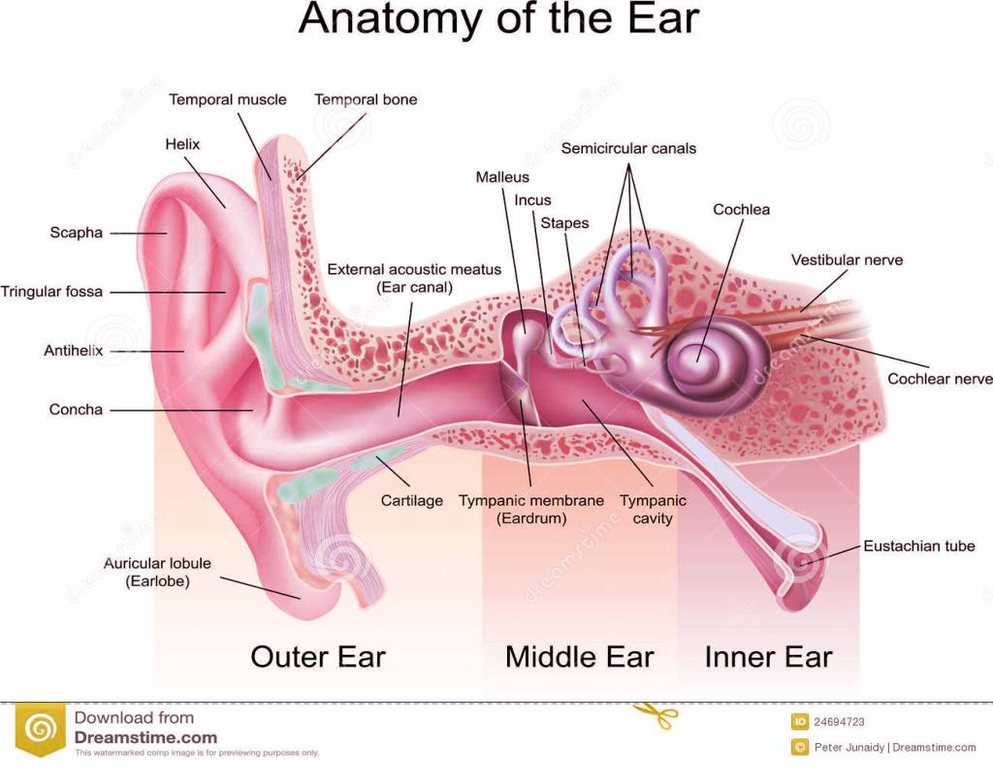
A brief description of the human ear along with a well-labelled diagram is given below for reference. Pinna/auricle is the outermost section of the ear. The external auditory canal links the exterior ear to the inner or the middle ear.
The middle ear is a chamber located within the petrous portion of the temporal bone. Structures within the middle ear amplify sound waves and transmit them to an appropriate portion of the internal ear. The internal ear contains the sensory organs for equilibrium (balance) and hearing. Figure 1. Ear structure Figure 2. Ear anatomy
The Structure of Human Ear Helix: It is the prominent outer rim of the external ear. Antihelix: It is the cartilage curve that is situated parallel to the helix. Crus of the Helix: It is the landmark of the outer ear, situated right above the pointy protrusion known as the tragus.