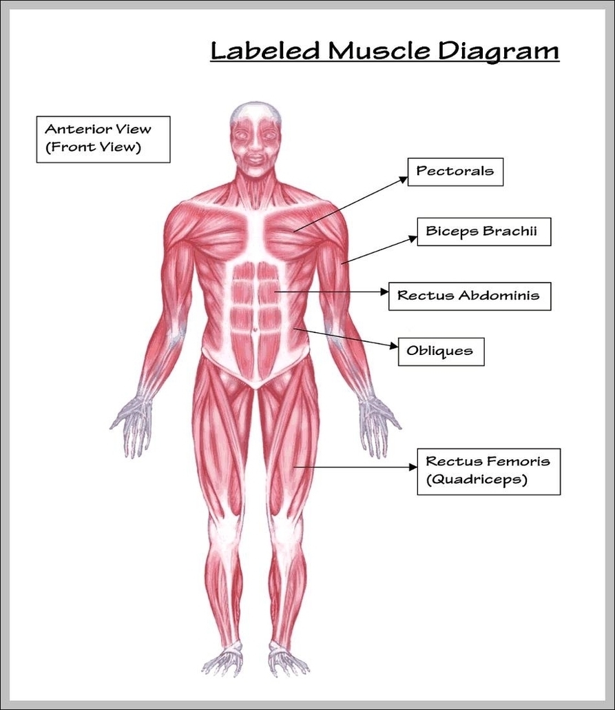
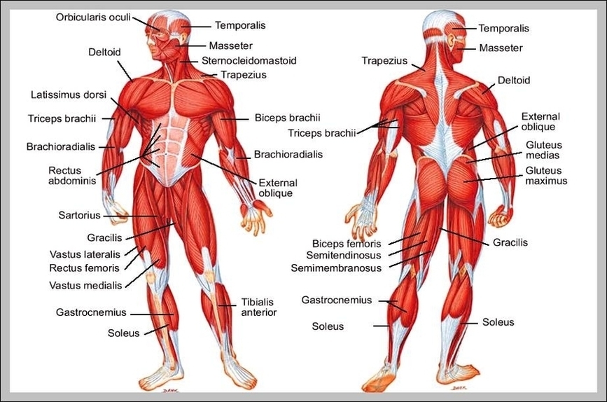
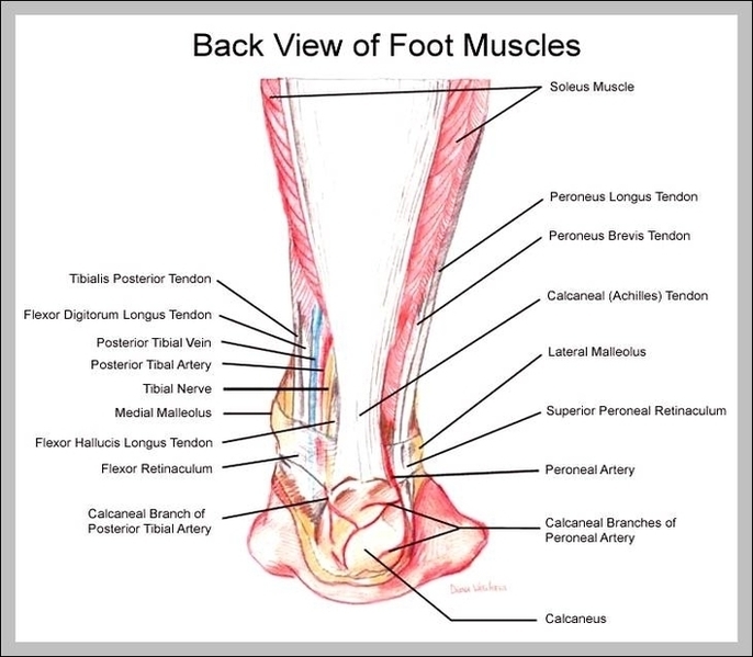
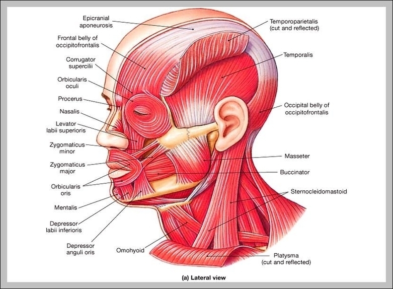
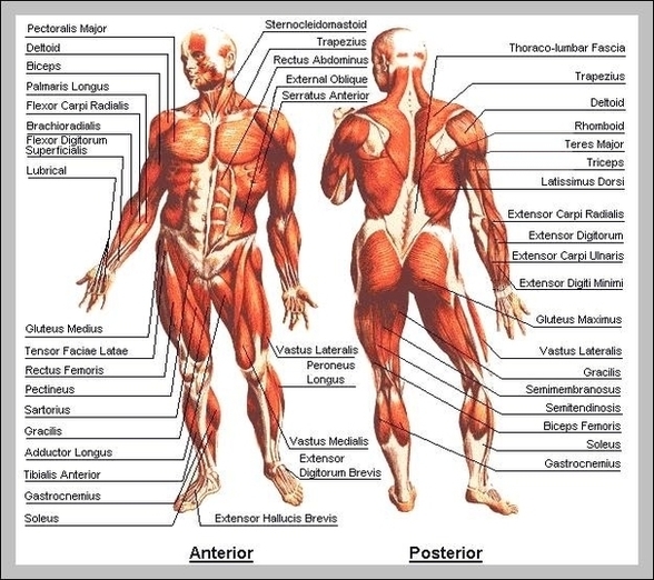
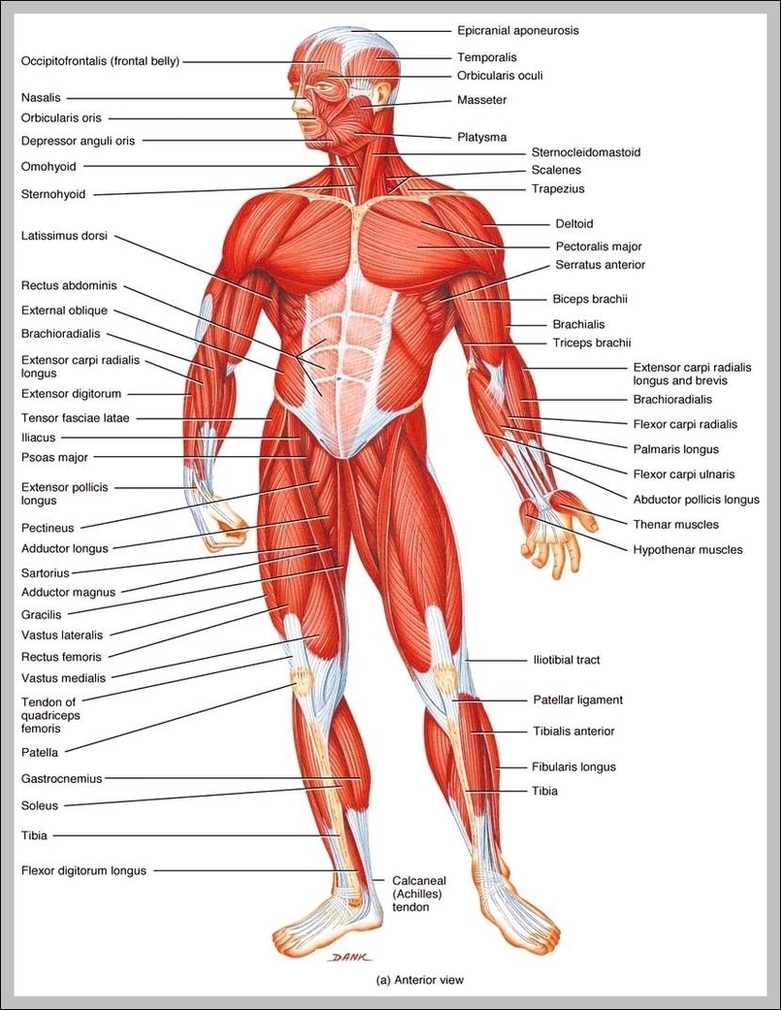
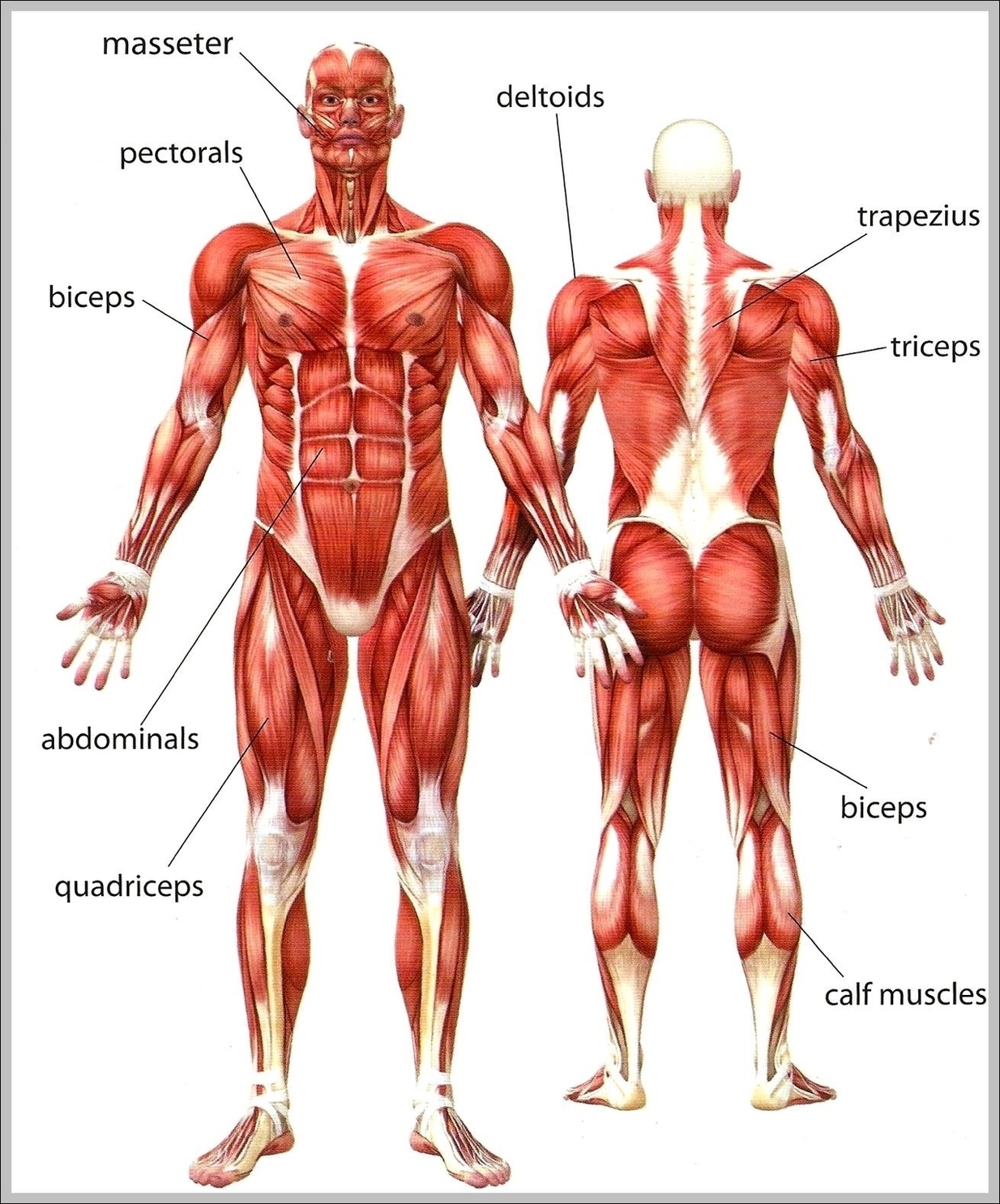
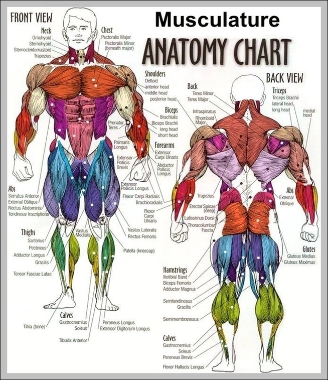
All Body Muscles Image
94,749 muscles body stock photos and images available, or search for oxygen or the human body to find more great stock photos and pictures.
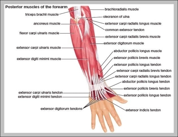
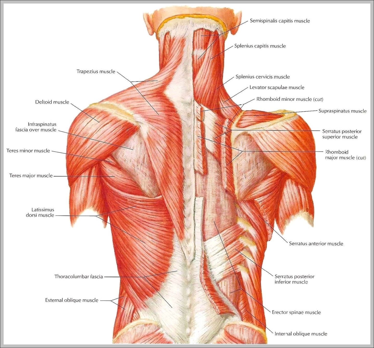
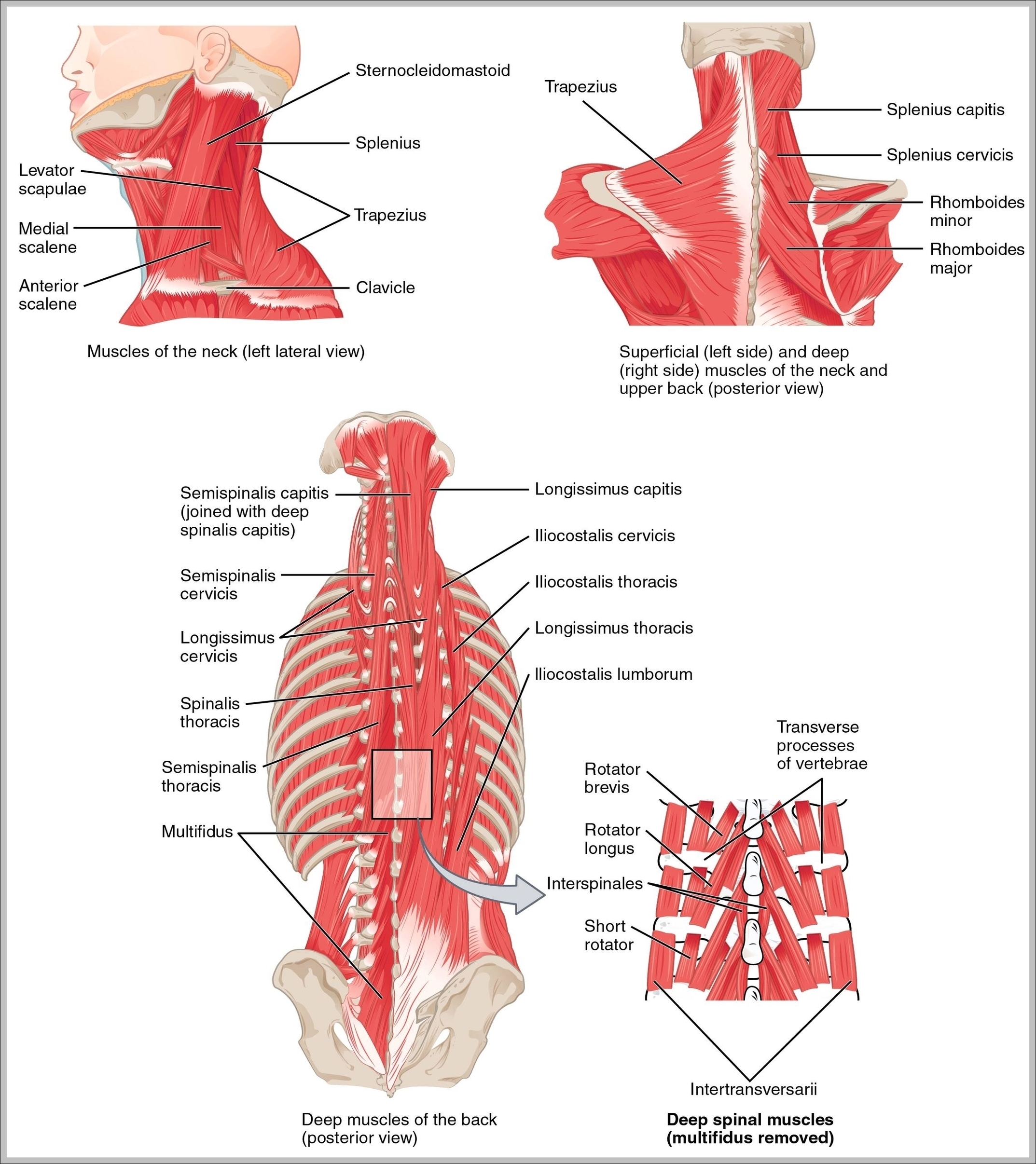
Forearms – Anatomy Muscles Rhomboid minor and rhomboid major, levator scapulae and latissimus dorsi muscles – didactic board of anatomy of human bony and muscular system, posterior view Abs – Anatomy Muscles Leg muscles of the man Human Body Organs (Lungs) 3D Triceps – Anatomy Muscles Hand muscle connection with brain
Forearms – Anatomy Muscles Rhomboid minor and rhomboid major, levator scapulae and latissimus dorsi muscles – didactic board of anatomy of human bony and muscular system, posterior view Abs – Anatomy Muscles Leg muscles of the man Human Body Organs (Lungs) 3D Triceps – Anatomy Muscles Hand muscle connection with brain