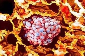
Diagram Cancerous Blood Cells
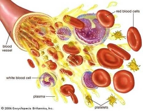
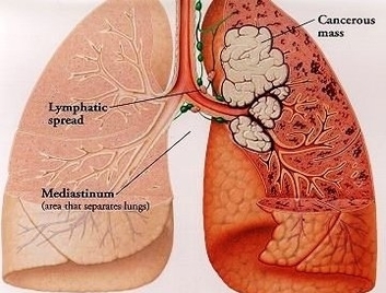
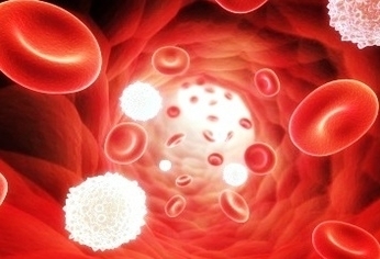
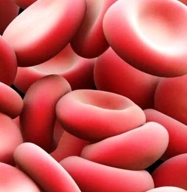
Cancer develops when cells in the body multiply out of control. Blood contains three kinds of cells: red cells, white cells, and platelets. Any of these kinds of cells can develop into cancer cells. So instead of a tumor (a clump of cancer cells) developing, such as in lung cancer, the tumor cells are spread throughout the blood system of the body.
Blood Cell Cancers. Cancer develops when cells in the body multiply out of control. Blood contains three kinds of cells: red cells, white cells, and platelets. Any of these kinds of cells can develop into cancer cells. So instead of a tumor (a clump of cancer cells) developing, such as in lung cancer, the tumor cells are spread throughout…
Cancer develops when cells in the body multiply out of control. Blood contains three kinds of cells: red cells, white cells, and platelets. Any of these kinds of cells can develop into cancer cells. So instead of a tumor (a clump of cancer cells) developing, such as in lung cancer, the tumor cells are spread throughout…